Fundus photography
| Fundus photography | |
|---|---|
| Intervention | |
| ICD-9-CM | 95.11 |


The left image (right eye) shows lighter areas close to larger vessels, which is regarded as a normal finding in younger people.
Fundus photography (also called fundography[1]) is the creation of a photograph of the interior surface of the eye, including the retina, optic disc, macula, and posterior pole (i.e. the fundus).[2][3]
Fundus photography is used by optometrists, ophthalmologists, and trained medical professionals for monitoring progression of a disease, diagnosis of a disease (combined with retinal angiography), or in screening programs and epidemiology.[4][5]
Compared to ophthalmoscopy, fundus photography generally needs a considerably larger instrument, but has the advantage of availing the image to be examined by a specialist at another location and/or time, as well as providing photo documentation for future reference. Modern fundus photographs generally recreate considerably larger areas of the fundus than what can be seen at any one time with handheld ophthalmoscopes.
Fundus camera

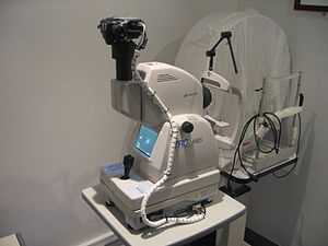
Fundus photography is performed by a fundus camera, which basically consists of a specialized low power microscope with an attached camera.
Optical principles
The optical design of fundus cameras is based on the principle of monocular indirect ophthalmoscopy.[2][3] A fundus camera provides an upright, magnified view of the fundus. A typical camera views 30 to 50° of retinal area, with a magnification of 2.5x, and allows some modification of this relationship through zoom or auxiliary lenses from 15°, which provides 5x magnification, to 140° with a wide angle lens, which minifies the image by half.[3] The optics of a fundus camera are similar to those of an indirect ophthalmoscope in that the observation and illumination systems follow dissimilar paths. The observation light is focused via a series of lenses through a doughnut shaped aperture, which then passes through a central aperture to form an annulus, before passing through the camera objective lens and through the cornea onto the retina.[6] The light reflected from the retina passes through the un-illuminated hole in the doughnut formed by the illumination system. As the light paths of the two systems are independent, there are minimal reflections of the light source captured in the formed image. The image forming rays continue towards the low powered telescopic eyepiece. When the button is pressed to take a picture, a mirror interrupts the path of the illumination system allow the light from the flash bulb to pass into the eye. Simultaneously, a mirror falls in front of the observation telescope, which redirects the light onto the capturing medium, whether it is film or a digital CCD. Because of the eye’s tendency to accommodate while looking though a telescope, it is imperative that the exiting vergence is parallel in order for an in focus image to be formed on the capturing medium.
Since the instruments are complex in design and difficult to manufacture to clinical standards, only a few manufacturers exist: Topcon, Zeiss, Canon, Nidek, Kowa, CSO and CenterVue.
Modes
Practical instruments for fundus photography perform the following modes of examination:
- Color, where the retina is illuminated by white light and examined in full color.
- Red-free, where the imaging light is filtered to remove red colors, improving contrast of vessels and other structures.
- Angiography, where the vessels are brought into high contrast by intravenous injection of a fluorescent dye. The retina is illuminated with an excitation color which fluoresces light of another color where the dye is present. By filtering to exclude the excitation color and pass the fluorescent color, a very high-contrast image of the vessels is produced. Shooting a timed sequence of photographs of the progression of the dye into the vessels reveals the flow dynamics and related pathologies. Specific methods include sodium fluorescein angiography (abbreviated FA or FAG) and indocyanine green (abbreviated ICG) angiography.
Indications
Fundus photography is used to detect and evaluate symptoms of retinal detachment or eye diseases such as glaucoma.
In patients with headaches, the finding of swollen optic discs, or papilledema, on fundus photography is a key sign, as this indicates raised intracranial pressure (ICP) which could be due to hydrocephalus, benign intracranial hypertension (aka pseudotumor cerebri) or brain tumor, amongst other conditions. Cupped optic discs are seen in glaucoma.
In patients with diabetes mellitus, regular fundus examinations (once every 6 months to 1 year) are important to screen for diabetic retinopathy as visual loss due to diabetes can be prevented by retinal laser treatment if retinopathy is spotted early.
In arterial hypertension, hypertensive changes of the retina closely mimic those in the brain, and may predict cerebrovascular accidents (strokes).
See also
- Dilated fundus examination
- Optical coherence tomography, commonly used for imaging the structure of the retina
Gallery
-

A close-up of the controls of a Topcon retinal camera
External links
- Ophthalmic Photographers' Society
- Fundus Photography
- "An objective focusing method for fundus photography."
References
- ↑ Lee, Y.; Lin, R. S.; Sung, F. C.; Yang, C.; Chien, K.; Chen, W.; Su, T.; Hsu, H.; Huang, Y. (2000). "Chin-Shan Community Cardiovascular Cohort in Taiwan-baseline data and five-year follow-up morbidity and mortality". Journal of clinical epidemiology 53 (8): 838–846. doi:10.1016/S0895-4356(00)00198-0. PMID 10942867.
- ↑ 2.0 2.1 Cassin, B. and Solomon, S. Dictionary of Eye Terminology. Gainsville, Florida: Triad Publishing Company, 1990.
- ↑ 3.0 3.1 3.2 Saine PJ. "Fundus Photography: What is a Fundus Camera?" Ophthalmic Photographers' Society. Accessed September 30, 2006.
- ↑ Adar SD, Klein R, Klein BE, Szpiro AA, Cotch MF, Wong TY, et al. 2010. Air Pollution and the microvasculature: a crosssectional assessment of in vivo retinal images in the population based multiethnic study of atherosclerosis (MESA). PLoS Med 7: e1000372.
- ↑ Louwies, T; Int Panis, L; Kicinski, M; De Boever, P; Nawrot, Tim S (2013). "Retinal Microvascular Responses to Short-Term Changes in Particulate Air Pollution in Healthy Adults". Environmental Health Perspectives. doi:10.1289/ehp.1205721.
- ↑ Saine PJ. "Fundus Photography: Fundus Camera Optics." Ophthalmic Photographers' Society. Accessed September 30, 2006.
| ||||||||||||||||||||||||||||||||||||||||||||||||
