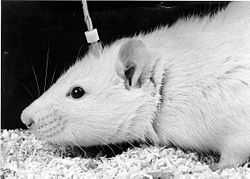
Electrical brain stimulation

Electrical brain stimulation (EBS), also referred to as focal brain stimulation (FBS), is a form of electrotherapy and technique used in research and clinical neurobiology to stimulate a neuron or neural network in the brain through the direct or indirect excitation of its cell membrane by using an electric current. It is used for research or for therapeutical purposes.
History
Electrical brain stimulation was first used in the first half of the 19th century by pioneering researchers such as Luigi Rolando [citation needed](1773–1831) and Pierre Flourens [citation needed](1794–1867), to study the brain localization of function, following the discovery by Italian physician Luigi Galvani (1737–1798) that nerves and muscles were electrically excitable. The stimulation of the surface of the cerebral cortex by using brain stimulation was used to investigate the motor cortex in animals by researchers such as Eduard Hitzig (1838–1907), Gustav Fritsch (1838–1927), David Ferrier (1842–1928) and Friedrich Goltz (1834–1902). The human cortex was also stimulated electrically by neurosurgeons and neurologists such as Robert Bartholow (1831–1904) and Fedor Krause (1857–1937).
In the following century, the technique was improved by the invention of the stereotactic method by British neurosurgeon pioneer Victor Horsley (1857–1916), and by the development of chronic electrode implants by Swiss neurophysiologist Walter Rudolf Hess (1881–1973), José Delgado (1915-2011) and others, by using electrodes manufactured by straight insulated wire that could be inserted deep into the brain of freely-behaving animals, such as cats and monkeys. This approach was used by James Olds (1922–1976) and colleagues to discover brain stimulation reward and the pleasure center. American-Canadian neurosurgeon Wilder Penfield (1891–1976) and colleagues at the Montreal Neurological Institute used extensively electrical stimulation of the brain cortex in awake neurosurgical patients to investigate the motor and sensory homunculus (the representation of the body in the brain cortex according to the distribution of motor and sensory territories).
Process
Two-photon excitation microscopy has shown that microstimulation activates neurons sparsely around the electrode even at low currents (as low as 10 μA) up to distances as far as four millimeters away. This happens without particularly selecting other neurons much nearer the electrode's tip. This is due to activation of neurons being determined by whether they do or do not have axons or dendrites that pass within a radius of 15 μm near the tip of the electrode. As the current is increased the volume around the tip that activates neuron axons and dendrites increases and with this the number of neurons activated. Activation is most likely to be due to direct depolarization rather than synaptic activation.[1]
Therapeutic applications

Examples of therapeutic EBS are:
- Cranial electrotherapy stimulation (CES)
- Deep brain stimulation (DBS)
- Transcranial direct current stimulation (tDCS)
- Electroconvulsive therapy (ECT)
- Functional electrical stimulation (FES)
- Magnetic seizure therapy (MST)
- Vagus nerve stimulation (VNS)
- Deep transcranial magnetic stimulation (Deep TMS)
Strong electric currents may cause a localized lesion in the nervous tissue, instead of a functional reversible stimulation. This property has been used for neurosurgical procedures in a variety of treatments, such as for Parkinson's disease, focal epilepsy and psychosurgery. Sometimes the same electrode is used to probe the brain for finding defective functions, before passing the lesioning current (electrocoagulation).
References
- ↑ Histed, MH; Bonin, V; Reid, RC. (2009). "Direct activation of sparse, distributed populations of cortical neurons by electrical microstimulation". Neuron 63 (4): 508–22. doi:10.1016/j.neuron.2009.07.016. PMC 2874753. PMID 19709632.