Contrast transfer function

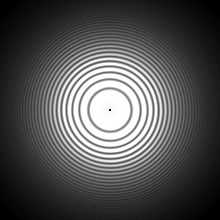
The contrast transfer function[1][2][3] is the equivalent of the optical transfer function in light that affects images collected in a transmission electron microscope. The contrast transfer function must be corrected in the images in order to obtain high resolution structures in three-dimensional electron microscopy, especially cryo-electron microscopy.
The oscillations of contrast transfer functions have the form (not including the envelope function):
where A is the amplitude contrast.[4] The amplitude contrast term can be converted into a phase shift, using the linear combination trigonometry rule:
where  . The function
  is defined as:
where r is the radius from the center of the image, Cs is the spherical aberration, λ is the wavelength of the electron beam (usually converted from the potential difference voltage) and z is the amount of defocus (using the convention that underfocus is negative and overfocus is positive)[4][5]
Furthermore, if the CTF is astigmatic, the defocus becomes a function of the angle θ where the astigmatic angle, θast given by:[6][7]
where  is the average defocus and
  is the difference between the maximal and minimal defocus in the CTF. Where the defocal difference is defined such that:
 or
or 
See also
- Optical transfer function
- Point spread function
- Contrast transfer function (CTF) correction
- Airy disk, different but similar phenomena in light
- Talk on the CTF by Henning Stahlberg
- CTF reading list
References
- ↑ Spence, John C. H. (1988 2nd ed) Experimental high-resolution electron microscopy (Oxford U. Press, NY) ISBN 0195054059.
- ↑ Ludwig Reimer (1997 4th ed) Transmission electron microscopy: Physics of image formation and microanalysis (Springer, Berlin) preview.
- ↑ Earl J. Kirkland (1998) Advanced computing in electron microscopy (Plenum Press, NY).
- ↑ 4.0 4.1 Malick, S.P. (2005). "ACE: Automated CTF Estimation". Ultramicroscopy 104 (1): 8–29. doi:10.1016/j.ultramic.2005.02.004.
- ↑ Maxim V. Sidorov. "What Is CTF (Contrast Transfer Function)?". ctfExplorer. Retrieved July 29, 2011.
- ↑ Mindell, J. A.; Grigorieff, N. (2003). "Accurate determination of local defocus and specimen tilt in electron microscopy". Journal of structural biology 142 (3): 334–347. PMID 12781660.
- ↑


