Ribosomal particles are denoted according to their sedimentation coefficients in Svedberg units. The 40S subunit is the small subunit of eukaryotic 80S ribosomes. It is structurally and functionally related to the 30S subunit of 70S prokaryotic ribosomes.[1][2][3][4][5] However, the 40S subunit is much larger than the prokaryotic 30S subunit and contains many additional protein segments, as well as rRNA expansion segments.
Function
The 40S subunit contains the decoding center which monitors the complementarity of tRNA and mRNA in protein translation. It is the target of most eukaryotic initiation factors and also interacts with the internal ribosome entry site of the hepatitis C virus.[6] More information can be found in the articles on the 80S eukaryotic ribosome, the ribosome in general and the article on protein translation.
Overall Structure
The shape of the small subunit can be subdivided into two large segments, the head and the body. Characteristic features of the body include the left and right feet, the shoulder and the platform. The head features a pointed protrusion reminiscent of a bird's beak. The mRNA binds in the cleft between the head and the body, and there are three binding sites for tRNA, the A-site, P-site and E-site (see article on protein translation for details).
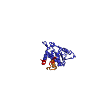
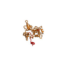
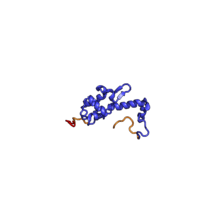
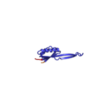
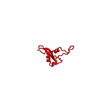
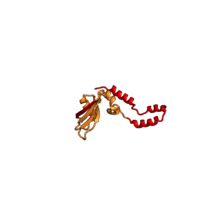
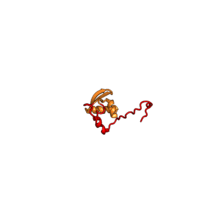
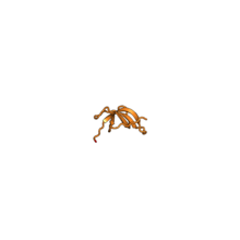
The core of the 40S subunit is formed by the 18S ribosomal RNA (abbreviated 18S rRNA), which is homologous to the prokaryotic 16S rRNA. This rRNA core is decorated with dozens of proteins. In the figure "Crystal Structure of the Eukaryotic 40S Ribosomal Subunit from T. thermophila", the ribosomal RNA core is represented as a grey tube and expansion segments are shown in red. Proteins which have homologs in eukaryotes, archaea and bacteria are shown as blue ribbons. Proteins shared only between eukaryotes and archaea are shown as orange ribbons and proteins specific to eukaryotes are shown as red ribbons.
| Crystal Structure of the Eukaryotic 40S Ribosomal Subunit from T. thermophila |
|---|
| 40S subunit viewed from the subunit interface side, PDB identifier 2XZM |
| 40S subunit viewed from the solvent-exposed side, PDB identifier 2XZM |
|
40S Ribosomal Proteins
The table "40S ribosomal proteins" shows the individual protein folds of the 40S subunit colored by conservation. Proteins which have homologs in eukaryotes, archaea and bacteria (EAB) are shown as blue ribbons. Proteins shared only between eukaryotes and archaea (EA) are shown as orange ribbons and proteins specific to eukaryotes (E) are shown as red ribbons. Eukaryote-specific extensions of conserved proteins, ranging from a few residues or loops to very long alpha helices and additional domains, are highlighted in red.[2] For a details, refer to the article on the eukaryotic ribosome. Historically, different nomenclatures have been used for ribosomal proteins. For instance, proteins have been numbered according to their migration properties in gel electrophoresis experiments. Therefore, different names may refer to homologous proteins from different organism, while identical names not necessarily denote homologous proteins. The table "40S ribosomal proteins" crossreferences the human ribosomal protein names with yeast, bacterial and archaeal homologs.[7] Further information can be found in the ribosomal protein gene database (RPG).[7]
40S ribosomal proteins
| Structure (Eukaryotic)[8] |
H. sapiens[7][9] |
Conservation[10] |
S. cerevisiae[11] |
Bacterial homolog (E. coli) |
Archaeal homolog |
| | RPSA | EAB | S0 | S2p | S2 |
| | RPS2 | EAB | S2 | S5p | S5p |
| | RPS3 | EAB | S3 | S3p | S3p |
| | RPS3A | EA | S1 | n/a | S3Ae |
| | RPS4 | EA | S4 | n/a | S4e |
| | RPS5 | EAB | S5 | S7p | S5p |
| | RPS6 | EA | S6 | n/a | S6e |
| | RPS7 | E | S7 | n/a | n/a |
| | RPS8 | EA | S8 | n/a | S8e |
| | RPS9 | EAB | S9 | S4p | S4p |
| | RPS10 | E | S10 | n/a | n/a |
| | RPS11 | EAB | S11 | S17p | S17p |
| | RPS12 | E | S12 | n/a | n/a |
| | RPS13 | EAB | S13 | S15p | S15p |
| | RPS14 | EAB | S14 | S11p | S11p |
| | RPS15 | EAB | S15 | S19p | S19p |
| | RPS15A | EAB | S22 | S8p | S8p |
| | RPS16 | EAB | S16 | S9p | S9p |
| | RPS17 | EA | S17 | n/a | S17e |
| | RPS18 | EAB | S18 | S13p | S13p |
| | RPS19 | EA | S19 | n/a | S19e |
| | RPS20 | EAB | S20 | S10p | S10p |
| | RPS21 | E | S21 | n/a | n/a |
| | RPS23 | EAB | S23 | S12p | S12p |
| | RPS24 | EA | S24 | n/a | S24e |
| | RPS25 | EA | S25 | n/a | S25e |
| | RPS26 | EA | S26 | n/a | S26e |
| | RPS27 | EA | S27 | n/a | S27e |
| | RPS27A | EA | S31 | n/a | S27ae |
| | RPS28 | EA | S28 | n/a | S28e |
| | RPS29 | EAB | S29 | S14p | S14p |
| | RPS30 | EA | S30 | n/a | S30e |
| | RACK1 | E | Asc1 | n/a | n/a |
External links
References
- ↑ 40S Ribosomal Subunits at the US National Library of Medicine Medical Subject Headings (MeSH)
- ↑ 2.0 2.1 Rabl J, Leibundgut M, Ataide SF, Haag A, Ban N. Crystal structure of the eukaryotic 40S ribosomal subunit in complex with initiation factor 1. Science. 2011 Feb 11;331(6018):730-6. doi: 10.1126/science.1198308. Epub 2010 Dec 23. PubMed PMID 21205638.
- ↑ Ben-Shem A, Garreau de Loubresse N, Melnikov S, Jenner L, Yusupova G, Yusupov M. The structure of the eukaryotic ribosome at 3.0 Ã
resolution. Science. 2011 Dec 16;334(6062):1524-9. doi: 10.1126/science.1212642. Epub 2011 Nov 17. PubMed PMID 22096102.
- ↑ Wimberly BT, Brodersen DE, Clemons WM Jr, Morgan-Warren RJ, Carter AP, Vonrhein C, Hartsch T, Ramakrishnan V. Structure of the 30S ribosomal subunit. Nature. 2000 Sep 21;407(6802):327-39. PubMed PMID 11014182.
- ↑ Schmeing TM, Ramakrishnan V. What recent ribosome structures have revealed about the mechanism of translation. Nature. 2009 Oct 29;461(7268):1234-42. doi:10.1038/nature08403. Epub 2009 Oct 18. Review. PubMed PMID 19838167.
- ↑ Lytle JR, Wu L, Robertson HD (August 2002). "Domains on the hepatitis C virus internal ribosome entry site for 40s subunit binding". RNA 8 (8): 1045–55. doi:10.1017/S1355838202029965. PMC 1370315. PMID 12212848.
- ↑ 7.0 7.1 7.2 Nakao A, Yoshihama M, Kenmochi N. RPG: the Ribosomal Protein Gene database. Nucleic Acids Res. 2004 Jan 1;32(Database issue):D168-70. PubMed PMID 14681386; PubMed Central PMCID: PMC308739.
- ↑ Structure of the T. thermophila,' proteins from the structures of the large subunit PDBS 417, 4A19 and small subunit PDB 2XZM
- ↑ Nomenclature according to the ribosomal protein gene database, applies to H. sapiens and T. thermophila
- ↑ EAB means conserved in eukaryotes, archaea and bacteria, EA means conserved in eukaryotes and archaea and E means eukaryote-specific protein
- ↑ Traditionally, ribosomal proteins were named according to their apparent molecular weight in gel electrophoresis, leading to different names for homologous proteins from different organisms. The RPG offers a unified nomenclature for ribosomal protein genes based on homology.
|
|---|
| | Prokaryotes (70S) | |
|---|
| | Eukaryotes (80S) |
|
|---|
|
B bsyn: dna (repl, cycl, reco, repr) · tscr (fact, tcrg, nucl, rnat, rept, ptts) · tltn (risu, pttl, nexn) · dnab, rnab/runp · stru (domn, 1°, 2°, 3°, 4°) |
|