Ovary
- "Ovaria" redirects here. This is also a proposed section and a synonym of Solanum.
| Ovary | |
|---|---|
 |
|
| Internal reproductive organs of human female | |
| Latin | ovarium |
| Gray's | subject #266 1254 |
| Artery | ovarian artery, uterine artery |
| Vein | ovarian vein |
| Nerve | ovarian plexus |
| Lymph | lumbar lymph nodes |
| MeSH | Ovary |
| Dorlands/Elsevier | Ovary |
The ovary is an ovum-producing reproductive organ, often found in pairs as part of the vertebrate female reproductive system. Ovaries in females are homologous to testes in males, in that they are both gonads and endocrine glands.
Contents |
Human anatomy
Ovaries are oval shaped and, in the human, measure approximately 3 cm x 1.5 cm x 1.5 cm (about the size of a Greek olive). The ovary (for a given side) is located in the lateral wall of the pelvis in a region called the ovarian fossa. The fossa usually lies beneath the external iliac artery and in front of the ureter and the internal iliac artery.
The ovaries aren't attached to the fallopian tubes but to the outer layer of the uterus via the ovarian ligaments. Usually each ovary takes turns releasing eggs every month; however, if there was a case where one ovary was absent or dysfunctional then the other ovary would continue providing eggs to be released.
Hormones
Ovaries secrete both estrogen and progesterone. Estrogen is responsible for the appearance of secondary sex characteristics of females at puberty and for the maturation and maintenance of the reproductive organs in their mature functional state. Progesterone functions with estrogen by promoting cyclic changes in the endometrium (it prepares the endometrium for pregnancy), as well as by helping maintain the endometrium in a healthy state during pregnancy.
Ligaments
In the human the paired ovaries lie within the pelvic cavity, on either side of the uterus, to which they are attached via a fibrous cord called the ovarian ligament. The ovaries are uncovered in the peritoneal cavity but are tethered to the body wall via the suspensory ligament of the ovary. The part of the broad ligament of the uterus that covers the ovary is known as the mesovarium. Thus, the ovary is the only organ in the human body which is totally invaginated into the peritonium, making it the only interperitoneal organ (not to be confused with intraperitoneal).
Extremities
There are two extremities to the ovary:
- The end to which the uterine tube attach is called the tubal extremity.
- The other extremity is called the uterine extremity. It points downward, and it is attached to the uterus via the ovarian ligament.
Histology
Cell Types
- Follicular cells flat epithelial cells that originate from surface epithelium covering the ovary
- granulosa cells - surrounding follicular cells have change from flat to cuboidal and proliferated to produce a stratified epithelium
- Gametes[1]

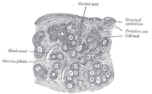
- The outermost layer is called the ovarian surface epithelium (previously known as the germinal epithelium).
- The tunica albuginea covers the cortex.
- The ovarian cortex consists of ovarian follicles and stroma in between them. Included in the follicles are the cumulus oophorus, membrana granulosa (and the granulosa cells inside it), corona radiata, zona pellucida, and primary oocyte. The zona pellucida, theca of follicle, antrum and liquor folliculi are also contained in the follicle. Also in the cortex is the corpus luteum derived from the follicles.
- The innermost layer is the ovarian medulla. It can be hard to distinguish between the cortex and medulla, but follicles are usually not found in the medulla.
In other animals
Ovaries of some kind are found in the female reproductive system of many animals that employ sexual reproduction, including invertebrates. However, they develop in a very different way in most invertebrates than they do in vertebrates, and are not truly homologous.[2]
Many of the features found in human ovaries are common to all vertebrates, including the presence of follicular cells, tunica albuginea, and so on. However, many species produce a far greater number of eggs during their lifetime than do humans, so that, in fish and amphibians, there may be hundreds, or even millions of fertile eggs present in the ovary at any given time. In these species, fresh eggs may be developing from the germinal epithelium throughout life. Corpora lutea are found only in mammals, and in some elasmobranch fish; in other species, the remnants of the follicle are quickly resorbed by the ovary. In birds, reptiles, and monotremes, the egg is relatively large, filling the follicle, and distorting the shape of the ovary at maturity.[2]
Amphibians and reptiles have no ovarian medulla; the central part of the ovary is a hollow, lymph-filled space. The ovary of teleosts is also often hollow, but in this case, the eggs are shed into the cavity, which opens into the oviduct.[2]
Although most normal female vertebrates have two ovaries, this is not the case in all species. In birds and platypuses, the right ovary never matures, so that only the left is functional. In some elasmobranchs, the reverse is true, with only the right ovary fully developing. In the primitive jawless fish, and some teleosts, there is only one ovary, formed by the fusion of the paired organs in the embryo.[2]
Cryopreservation
Cryopreservation of ovarian tissue is of interest to women who want to preserve their reproductive function beyond the natural limit, or whose reproductive potential is threatened by cancer therapy,[3] for example in hematologic malignancies or breast cancer.[4] The procedure is to take a part of the ovary and carry out slow freezing before storing it in liquid nitrogen whilst therapy is undertaken. Tissue can then be thawed and implanted near the fallopian, either orthotopic (on the natural location) or heterotopic (on the abdominal wall),[4] where it starts to produce new eggs, allowing normal conception to take place. [5] A study of 60 procedures concluded that ovarian tissue harvesting appears to be safe.[4] The ovarian tissue may also be transplanted into mice that are immunocompromised (SCID mice) to avoid graft rejection, and tissue can be harvested later when mature follicles have developed.[6]
Additional images
 Uterus and uterine tubes |
 Organs of the female reproductive system. |
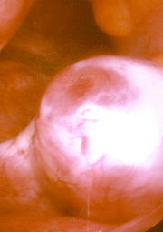
 Ovary |
 An ovary about to release an egg. |
 Vessels of the uterus and its appendages, rear view. |
 Broad ligament of adult, showing epoöphoron. |
 Uterus and right broad ligament, seen from behind. |
 Female pelvis and its contents, seen from above and in front. |
 Arteries of the female reproductive tract: uterine artery, ovarian artery and vaginal arteries. |
References
- ↑ Langman's Medical Embryology, Lippincott Williams & Wilkins, 10th ed, 2006
- ↑ 2.0 2.1 2.2 2.3 Romer, Alfred Sherwood; Parsons, Thomas S. (1977). The Vertebrate Body. Philadelphia, PA: Holt-Saunders International. pp. 383–385. ISBN 0-03-910284-X.
- ↑ Isachenko V, Lapidus I, Isachenko E, et al. (2009). "Human ovarian tissue vitrification versus conventional freezing: morphological, endocrinological, and molecular biological evaluation.". Reproduction 138: 319–27. PMID 19439559.
- ↑ 4.0 4.1 4.2 Oktay K, Oktem O (November 2008). "Ovarian cryopreservation and transplantation for fertility preservation for medical indications: report of an ongoing experience". Fertil. Steril.. doi:10.1016/j.fertnstert.2008.10.006. PMID 19013568.
- ↑ Livebirth after orthotopic transplantation of cryopreserved ovarian tissue The Lancet, Sep 24, 2004
- ↑ Lan C, Xiao W, Xiao-Hui D, Chun-Yan H, Hong-Ling Y (December 2008). "Tissue culture before transplantation of frozen-thawed human fetal ovarian tissue into immunodeficient mice". Fertil. Steril.. doi:10.1016/j.fertnstert.2008.10.020. PMID 19108826.
External links
- [1] From the American Medical Association
- [2] Merck Online Medical Library: Female Reproductive System
See also
|
|||||||||||||||||||||||||||||||||||||||||||||||||||||||||||||
|
||||||||||||||||||||||||||||||||||||||||||||||||||||||||||||||||||
|
||||||||||||||||||||||||||||||||||||||||||||||