Artificial respiration
Artificial respiration is the act of simulating respiration, which provides for the overall exchange of gases in the body by pulmonary ventilation, external respiration and internal respiration.[1] This means providing air for a person who is not breathing or is not making sufficient respiratory effort on their own[2] (although it must be used on a patient with a beating heart or as part of cardiopulmonary resuscitation to achieve the internal respiration).
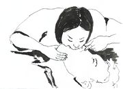
Pulmonary ventilation (and hence external respiration) is achieved through manual insufflation of the lungs either by the rescuer blowing into the patient's lungs, or by using a mechanical device to do so. This method of insufflation has been proved more effective than methods which involve mechanical manipulation of the patients chest or arms, such as the Silvester method.[3] It is also known as Expired Air Resuscitation (EAR), Expired Air Ventilation (EAV), mouth-to-mouth resuscitation, rescue breathing or colloquially the kiss of life.
Artificial respiration is a part of most protocols for performing cardiopulmonary resuscitation (CPR)[4][5] making it an essential skill for first aid. In some situations, artificial respiration is also performed separately, for instance in near-drowning and opiate overdoses. The performance of artificial respiration in its own is now limited in most protocols to health professionals, whereas lay first aiders are advised to undertake full CPR in any case where the patient is not breathing sufficiently.
Mechanical ventilation involves the use of a mechanical ventilator to move air in and out of the lungs when an individual is unable to breathe on his or her own, for example during surgery with general anesthesia or when an individual is in a coma.
Contents |
Insufflations

Insufflation, also known as 'rescue breaths' or 'ventilations', is the act of mechanically forcing air into a patient's respiratory system. This can be achieved via a number of methods, which will depend on the situation and equipment available. All methods require good airway management to perform, which ensures that the method is effective. These methods include:
- Mouth to mouth - This involves the rescuer making a seal between their mouth and the patient's mouth and 'blowing', to pass air into the patient's body
- Mouth to nose - In some instances, the rescuer may need or wish to form a seal with the patient's nose. Typical reasons for this include maxillofacial injuries, performing the procedure in water or the remains of vomit in the mouth
- Mouth to mouth and nose - Used on infants (usually up to around 1 year old), as this forms the most effective seal
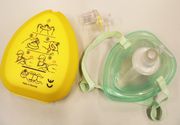
- Mouth to mask – Most organisations recommend the use of some sort of barrier between rescuer and patient to reduce cross infection risk. One popular type is the 'pocket mask'. This may be able to provide higher tidal volumes than a Bag Valve Mask.[6]
- Bag valve mask (BVM) - This is a simple device manually operated by the rescuer, which involves squeezing a bag to expel air into the patient.
- Mechanical resuscitator - An electric unit designed to breathe for the patient
Adjuncts to insufflation

Most training organisations recommend that in any of the methods involving mouth to patient, that a protective barrier is used, to minimise the possibility of cross infection (in either direction).[7]
Barriers available include pocket masks and keyring-sized face shields. These barriers are an example of Personal Protective Equipment to guard the face against splashing, spraying or splattering of blood or other potentially infectious materials.
These barriers should provide a one-way filter valve which lets the air from the rescuer deliver to the patient while any substances from the patient (e.g. vomit, blood) cannot reach the rescuer. Many adjuncts are single use, though if they are multi use, after use of the adjunct, the mask must be cleaned and autoclaved and the filter replaced.
The CPR mask is the preferred method of ventilating a patient when only one rescuer is available. Many feature 18mm inlets to support supplemental oxygen, which increases the oxygen being delivered from the approximate 17% available in the expired air of the rescuer to around 40-50%.
Tracheal intubation is often used for short term mechanical ventilation. A tube is inserted through the nose (nasotracheal intubation) or mouth (orotracheal intubation) and advanced into the trachea. In most cases tubes with inflatable cuffs are used for protection against leakage and aspiration. Intubation with a cuffed tube is thought to provide the best protection against aspiration. Tracheal tubes inevitably cause pain and coughing. Therefore, unless a patient is unconscious or anesthetized for other reasons, sedative drugs are usually given to provide tolerance of the tube. Other disadvantages of tracheal intubation include damage to the mucosal lining of the nasopharynx or oropharynx and subglottic stenosis.
In an emergency a Cricothyrotomy can be used by health care professionals, where an airway is inserted through a surgical opening in the cricothyroid membrane. This is similar to a tracheostomy but a cricothyrotomy is reserved for emergency access. This is usually only used when there is a complete blockage of the pharynx or there is massive maxillofacial injury, preventing other adjunts being used.[8]
Efficiency of mouth to patient insufflation
Normal atmospheric air contains approximately 21% oxygen when created in. After gaseous exchange has taken place in the lungs, with waste products (notably carbon dioxide) moved from the bloodstream to the lungs, the air being exhaled by humans normally contains around 17% oxygen. This means that the human body utilises only around 19% of the oxygen inhaled, leaving over 80% of the oxygen available in the exhalatory breath.[9]
This means that there is more than enough residual oxygen to be used in the lungs of the patient, which then crosses the cell membrane to form oxyhemoglobin.
Oxygen

The efficiency of artificial respiration can be greatly increased by the simultaneous use of oxygen therapy. The amount of oxygen available to the patient in mouth to mouth is around 16%. If this is done through a pocket mask with an oxygen flow, this increases to 40% oxygen. If a Bag Valve Mask or mechanical respirator is used with an oxygen supply, this rises to 99% oxygen. The greater the oxygen concentration, the more efficient the gaseous exchange will be in the lungs.
See also
- mechanical ventilation for a detailed discussion from the medical perspective.
- cardiopulmonary resuscitation
- medical emergency
References
- ↑ Tortora, Gerard J; Derrickson, Bryan (2006). Principles of Anatomy and Physiology. John Wiley & Sons Inc..
- ↑ "Artificial Respiration". Encyclopaedia Britannica. http://www.britannica.com/eb/article-9009713/artificial-respiration. Retrieved 2007-06-15.
- ↑ "Artificial Respiration". Microsoft Encarta Online Encyclopedia 2007. Archived from the original on 2009-10-31. http://www.webcitation.org/5kwKiCVG5. Retrieved 2007-06-15.
- ↑ "Decisions about cardiopulmonary resuscitation model information leafler". British Medical Association. July 2002. http://www.bma.org.uk/ap.nsf/Content/cprleaflet. Retrieved 2007-06-15.
- ↑ "Overview of CPR". American Heart Association. 2005. http://circ.ahajournals.org/cgi/content/full/112/24_suppl/IV-12. Retrieved 2007-06-15.
- ↑ Dworkin, Gerald M (Winter 1987). "Mouth to Mouth rescue breathing and comparisons of personal resuscitation masks". Rescue Squad Quarterly. http://www.lifesaving.com/issues/articles/30mouth_to_mouth.html. Retrieved 2007-06-15.
- ↑ "Emergency Cardiovascular Care Revisions for the professional rescuer" (DOC). American Red Cross. http://www.redcross.org/services/hss/resources/eccpr.doc. Retrieved 2007-06-15.
- ↑ Carley SD, Gwinnutt C, Butler J, Sammy I, Driscoll P. (March 2002). "Rapid sequence induction in the emergency department: a strategy for failure". Emergency Medicine Journal 19 (2): 109–113. doi:10.1136/emj.19.2.109. PMID 11904254. PMC PMC1725832. http://emj.bmjjournals.com/cgi/content/full/19/2/109. Retrieved 2007-05-19.
- ↑ "Physical Intervention: Life Support (Rescue Breathing)". http://www.doitnow.org/pages/208/208-5.html. Retrieved December 29, 2005.