Uterus
| Uterus | |
|---|---|
 |
|
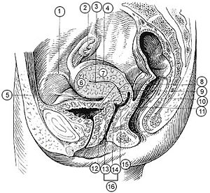
| Female internal reproductive anatomy | |
 |
|
| 1. Round ligament 2. Uterus 3. Uterine cavity 4. Intestinal surface of Uterus 5. Versical surface(toward bladder) 6. Fundus of uterus 7. Body of uterus 8. Palmate folds of cervical canal 9. Cervical canal 10. Posterior lip 11. Cervical os (external) 12. Isthmus of uterus 13. Supravaginal portion of cervix 14. Vaginal portion of cervix 15. Anterior lip 16. Cervix |
|
| Gray's | subject #268 1258 |
| Artery | ovarian artery, uterine artery, helicine branches of uterine artery |
| Vein | uterine veins |
| Lymph | body and cervix to internal iliac lymph nodes, fundus to superficial inguinal lymph nodes |
| Precursor | Müllerian duct |
| MeSH | Uterus |
The uterus (Latin word for womb) is a major female reproductive organ of most mammals, including humans. It is within the uterus that the fetus develops during gestation. The term uterus is used exclusively within the medical and related professions; some lay persons use the less formal term, womb. The plural of uterus is uteruses or uteri.
One end, the cervix, opens into the vagina; the other is connected on both sides to the Fallopian tubes.
Contents |
Function
The main function of the uterus is to accept a fertilized ovum which becomes implanted into the endometrium, and derives nourishment from blood vessels which develop exclusively for this purpose. The fertilized ovum becomes an embryo, develops into a fetus and gestates until childbirth. Due to anatomical barriers such as the pelvis, the uterus is pushed partially into the abdomen due to its expansion during pregnancy. Even during pregnancy the mass of a human uterus amounts to only about a kilogram (2.2 pounds).
Forms in mammals
In mammals, the four main forms in which it is found are:
- Bipartite
- Found in ungulates (deer, moose, elk etc.), and in carnivores (cats, and dogs).
- Bicornuate
- Found in pigs.
- Simplex
- Found in humans, other primates and horses.
- Duplex
- Found in rodents (such as mice, rats and guinea pigs), marsupials and lagomorpha (rabbits and hares).
Anatomy
The uterus is located inside the pelvis immediately dorsal (and usually somewhat rostral) to the urinary bladder and ventral to the rectum. Outside of pregnancy, its size in humans is several centimeters in diameter. The uterus is a pear shaped muscular organ which can be divided anatomically into four segments: The fundus, corpus, cervix and the internal os.
Regions
From outside to inside, the path to the uterus is as follows:
- Vulva
- Vagina
- Cervix uteri - "neck of uterus"
- External orifice of the uterus
- Canal of the cervix
- Internal orifice of the uterus
- corpus uteri - "Body of uterus"
- Cavity of the body of the uterus
- Fundus (uterus)
Layers
The layers, from innermost to outermost, are as follows:
- Endometrium
- The lining of the uterine cavity is called the "endometrium." In most mammals, including humans, the endometrium builds a lining periodically which, if no pregnancy occurs, is shed or reabsorbed. Shedding of the endometrial lining in humans is responsible for menstrual bleeding (known colloquially as a woman's "period") throughout the fertile years of a female and for some time beyond. In other mammals there may be cycles set as widely apart as six months or as frequently as a few days.
- Myometrium
- The uterus mostly consists of smooth muscle, known as "myometrium." The innermost layer of myometrium is known as the junctional zone, which becomes thickened in adenomyosis.
- Perimetrium
- The loose surrounding tissue is called the "perimetrium."
- Peritoneum
- The uterus is surrounded by "peritoneum."
Support
The uterus is primarily supported by the pelvic diaphragm and the urogenital diaphragm. Secondarily, it is supported by ligaments and the peritoneum (broad ligament of uterus) [1]
Major ligaments
It is held in place by several peritoneal ligaments, of which the following are the most important (there are two of each):
| Name | From | To |
|---|---|---|
| uterosacral ligament | the posterior cervix | the sacrum of pelvis |
| cardinal ligaments | the side of the cervix | the ischial spines |
| pubocervical ligament [1] |
Other named ligaments near the uterus, i.e. the broad ligament, the round ligament, the suspensory ligament of the ovary, the infundibulopelvic ligament, have no role in the support of the uterus.
Position
Under normal circumstances the uterus is both "anteflexed" and "anteverted." The meaning of these terms are described below:
| Distinction | More common | Less common |
|---|---|---|
| Position tipped | "anteverted": tipped forward | "retroverted": tipped backwards |
| Position of fundus | "anteflexed": the fundus is pointing forward relative to the cervix | "retroflexed": the fundus is pointing backwards |
Development
The bilateral Müllerian ducts form during early fetal life. In males, MIF secreted from the testes leads to their regression. In females these ducts give rise to the Fallopian tubes and the uterus. In humans the lower segments of the two ducts fuse to form a single uterus, however, in cases of uterine malformations this development may be disturbed. The different uterine forms in various mammals are due to various degrees of fusion of the two Müllerian ducts.
Pathology
Some pathological states include:
- Prolapse of the uterus
- Carcinoma of the cervix – malignant neoplasm
- Carcinoma of the uterus – malignant neoplasm
- Fibroids – benign neoplasms
- Adenomyosis – ectopic growth of endometrial tissue within the myometrium
- Pyometra – infection of the uterus, most commonly seen in dogs
- Uterine malformations mainly congenital malformations including Uterine Didelphys, bicornuate uterus and septate uterus. It also includes congenital absence of the uterus Rokitansky Syndrome
Additional images
References
- ↑ 1.0 1.1 The Pelvis University College Cork
See also
External links
- Gray's s268
- Illustration at wku.edu
- SUNY Labs 43:01-0102 - "The Female Pelvis: Organs in the Female Pelvis in situ"
|
|||||||||||||||||||||||||||||||||||||||