Urticaria
| Urticaria Classification and external resources |
|
 |
|
|---|---|
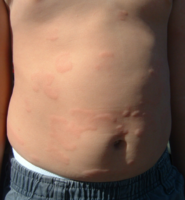
| Urticaria on foot, with visible wheals | |
| ICD-10 | L50. |
| ICD-9 | 708 |
| DiseasesDB | 13606 |
| MedlinePlus | 000845 |
| eMedicine | topic list |
| MeSH | D014581 |
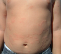
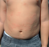
Urticaria (or hives) are a kind of skin rash notable for red, raised, itchy bumps. Hives are frequently caused by allergic reactions, however there are multiple non-allergic causes. For example, most cases of hives lasting less than 6 weeks (acute urticaria) are the result of an allergic trigger. Chronic urticaria (hives lasting longer than 6 weeks) are rarely due to an allergy. The majority of patients with chronic hives have an unknown (idiopathic) cause. Perhaps as many as 30-40% of patients with chronic idiopathic urticaria will, in fact, have an autoimmune cause. Acute viral infection is another common cause of acute urticaria (viral exanthem). Less common causes of hives include friction, pressure, temperature extremes, exercise, and sunlight. It has been said that hives are more common in those with fair skin.
Weals (raised areas surrounded by a red base) from urticaria can appear anywhere on the surface of the skin. Whether the trigger is allergic or non-allergic, there is a complex release of inflammatory mediators, including histamine from cutaneous mast cells, resulting in fluid leakage from superficial blood vessels. Weals may be pinpoint in size, or several inches in diameter. Angioedema is a related condition (also from allergic and non-allergic causes), though fluid leakage is from much deeper blood vessels. Individual hives that are painful, last >24 hours, or leave a bruise as they heal are more likely to be a more serious condition called urticaria pigmentosa. Hives caused by stroking the skin (often linear in appearance) is due to a benign condition called dermatographism.
Contents |
Pathophysiology

The skin lesions of urticarial disease are caused by an inflammatory reaction in the skin, causing leakage of capillaries in the dermis, and resulting in an edema which persists until the interstitial fluid is absorbed into the surrounding cells.
Urticaria are caused by the release of histamine and other mediators of inflammation (cytokines) from cells in the skin. This process can be the result of an allergic or non-allergic reaction, differing in the eliciting mechanism of histamine release.

- Allergic urticaria
- Histamine and other pro-inflammatory substances are released from mast cells in the skin and tissues in response to the binding of allergen-bound IgE antibodies to high affinity cell surface receptors. Basophils and other inflammatory cells are also seen to release histamine and other mediators, and are thought to play an important role, especially in chronic urticarial diseases.
- Autoimmune urticaria
- In the past decade, it has been noted that many cases of chronic idiopathic urticaria are the result of an autoimmune trigger. For example, roughly one third of patients with chronic urticaria spontaneously develop auto-antibodies directed at the receptor FcεRI located on skin mast cells. Chronic stimulation of this receptor leads to chronic hives. Patients often have other autoimmune conditions such as autoimmune thyroiditis.
- Infectious
- Hive-like rashes commonly accompany viral illnesses, such as the common cold. They usually appear 3-5 days after the cold has started, and may even appear a few days after the cold has resolved.
- Non-allergic urticaria
- Mechanisms other than allergen-antibody interactions are known to cause histamine release from mast cells. Many drugs, for example morphine, can induce direct histamine release not involving any immunoglobulin molecule. Also, a diverse group of signaling substances called neuropeptides have been found to be involved in emotionally induced urticaria. Dominantly inherited cutaneous and neurocutaneous porphyrias (porphyria cutanea tarda, hereditary coproporphyria, variegate porphyria and erythropoietic protoporphyria) have been associated with solar urticaria. The occurrence of drug-induced solar urticaria may be associated with porphyrias. This may be caused by IgG binding not IgE.
Differential diagnosis
The rash that develops from poison ivy, poison oak, and poison sumac contact is commonly mistaken for urticaria. This rash is caused by contact with urushiol and results in a form of contact dermatitis called Urushiol-induced contact dermatitis. Urushiol is spread by contact, but can be washed off with a strong grease/oil dissolving detergent and cool water and rubbing ointments.
Types
- Acute urticaria usually show up a few minutes after contact with the allergen and can last a few hours to several weeks. Food allergic reactions often fit in this category. The most common food allergies in adults are shellfish and nuts. The most common food allergies in chidren are shellfish, nuts, peaunts, eggs, wheat, and soy. It is uncommon for patients to have more than 2 true food allergies. A less common cause is exposure to certain bacteria, such as streptococcus or possibly Helicobacter pylori. [1] In these cases, the hives may be exacerbated by other factors, such as those listed under Physical Urticarias below.
- Chronic urticaria refers to hives that persists for 6 weeks or more. There are no visual differences between acute and chronic urticaria. Some of the more severe chronic cases have lasted more than 20 years. A survey indicated that chronic urticaria lasted a year or more in more than 50% of sufferers and 20 years or more in 20% of them. Of course this does mean that in almost half the people it clears up within a year and in 80% it clears up within 20 years or less.[2]
- Drug-induced urticaria has been known to result in severe cardiorespiratory failure. The anti-diabetic sulphonylurea glimepiride (trade name Amaryl®), in particular, has been documented to induce allergic reactions manifesting as urticaria. Other cases include dextroamphetamine[3], aspirin, penicillin, clotrimazole, sulfonamides and anticonvulsants.
- Physical urticarias are often categorized into the following.
- Aquagenic: Reaction to water (exceedingly rare)
- Cholinergic: Reaction to body heat, such as when exercising or after a hot shower
- Cold (Chronic cold urticaria): Reaction to cold, such as ice, cold air or water - worse with sudden change in temperature
- Delayed Pressure: Reaction to standing for long periods, bra-straps, elastic bands on undergarments, belts
- Dermatographic: Reaction when skin is scratched (very common)
- Heat: Reaction to hot food or objects (rare)
- Solar: Reaction to direct sunlight (rare, though more common in those with fair skin)
- Vibration: Reaction to vibration (rare)
- Adrenergic: Reaction to adrenaline / noradrenaline (extremely rare)
Related conditions
Angioedema is similar to urticaria,[4] but in angioedema, the swelling occurs in a lower layer of the dermis than it does in urticaria[5], as well as in the subcutis. This swelling can occur around the mouth, in the throat, in the abdomen, or in other locations. Urticaria and angioedema sometimes occur together in response to an allergen and is a concern in severe cases as angioedema of the throat can be fatal.
Treatment and management
Chronic urticaria can be difficult to treat. There are no guaranteed treatments or means of controlling attacks, and some sub-populations are treatment resistant, with medications spontaneously losing their effectiveness and requiring new medications to control attacks. It can be difficult to determine appropriate medications since some, such as loratadine, require a day or two to build up to effective levels, and since the condition is intermittent and outbreaks typically clear up without any treatment.
Most treatment plans for urticaria involve being aware of one's triggers, but this can be difficult since there are several different forms of urticaria and people often exhibit more than one type. Also, since symptoms are often idiopathic (unknown reason) there might not be any clear trigger. If one's triggers can be identified then outbreaks can often be managed by limiting one's exposure to these situations.
Histamine antagonists
Drug treatment is typically in the form of Antihistamines such as diphenhydramine, hydroxyzine, cetirizine and other H1 receptor antagonists.[6] These are taken on a regular basis to protective effect, lessening or halting attacks. While the disease is obviously physiological in origin, psychological treatments such as stress management can sometimes lessen severity and occurrence. Additionally, methods similar to psychological pain management can be used to shift focus away from the discomfort and itchiness during an attack.
The H2-receptor antagonists such as cimetidine and ranitidine may help control symptoms either prophylactically or by lessening symptoms during an attack.[7] When taken in combination with a H1 antagonist it has been shown to have a synergistic effect which is more effective than either treatment alone. The use of ranitidine (or other H2 antagonist) for urticaria is considered an off-label use, since these drugs are primarily used for the treatment of peptic ulcer disease and gastroesophageal reflux disease.
Other
Tricyclic antidepressants such as doxepin, also are often potent H1 and H2 antagonists and may have a role in therapy, although side effects limit their use. For very severe outbreaks, an oral corticosteroid such as Prednisone is sometimes prescribed. However this form of treatment is controversial because of the extensive side effects common with corticosteroids and as such is not a recommended long-term treatment option.
As of 2008 an [Australian] company is performing clinical trials with an analogue of alpha-melanocyte-stimulating hormone called melanotan (known by the International Nonproprietary Name afamelanotide, formerly CUV1647)[8] for the treatment of solar urticaria,[9][10] a type of urticaria that develops in response to exposure to specific wavelengths of light.[11]
Dietary
Children with intermittent or recurrent urticaria-angiodema were fed 7 food-additives: tartrazine (E102), sunset yellow (E110), erythrosine (E127), annatto (E160b), sodium benzoate (E211), acetyl-salicylic acid (ASA) and aspartame. Reactions to the food additives were common: E110 = 64%, E160b = 60%, E211: 57%; E102 = 50%, aspartame = 48%, E127 = 35%, ASA = 12%. The authors suggest that food additive intolerance is frequent in children with recurrent or intermittent urticaria-angiodema, and that aspartame may contribute directly to urticaria-angiodema in childhood.[12]
See also
- Anti-itch drug
- Chronic cold urticaria
- Urticaria pigmentosa
- Cholinergic urticaria
|
References
- ↑ Tebbe B, Geilen CC, Schulzke JD, Bojarski C, Radenhausen M, Orfanos CE (April 1996). "Helicobacter pylori infection and chronic urticaria". J. Am. Acad. Dermatol. 34 (4): 685–6. PMID 8601663. http://linkinghub.elsevier.com/retrieve/pii/S0190-9622(96)80086-7.
- ↑ Champion RH, Roberts SO, Carpenter RG, Roger JH (1969). "Urticaria and angio-oedema. A review of 554 patients". Br. J. Dermatol. 81 (8): 588–97. PMID 5801331.
- ↑ "Prescribing Information Dexedrine". GlaxoSmithKline (June 2006).
- ↑ angioedema at Dorland's Medical Dictionary
- ↑ "Hives (Urticaria and Angioedema)" (2006-03-01). Retrieved on 2007-08-24.
- ↑ Greaves MW, Tan KT (2007). "Chronic Urticaria: Recent Advances". Clin Rev Allergy Immunol 33 (1-2): 134–143. doi:. PMID 18094952.
- ↑ Lee EE, Maibach HI (2001). "Treatment of urticaria. An evidence-based evaluation of antihistamines". Am J Clin Dermatol 2 (1): 27–32. PMID 11702618.
- ↑ "World Health Organisation assigns CUV1647 generic name" (PDF). Clinuvel (2008). Retrieved on 2008-06-17.
- ↑ McDonald, Kate (2007-04-13). "Tackling skin cancer in organ transplant patients". Australian Life Scientist. Retrieved on 2007-12-24.
- ↑ "Clinuvel gets green light". Biotechnews.com.au (LifeScientist) (2007-06-11). Retrieved on 2008-06-13.
- ↑ Baron, ED; Taylor, CR (2007-03-29). "Urticaria, Solar". WebMD. Retrieved on 2007-12-26.
- ↑ de Martino M, Peruzzi M, Galli L, Lega L, Zammarchi E, Vierucci A (1992). "Food-additive intolerance and its correlation with atopy in children with recurrent or intermittent urticaria-angioedema". Pediatric Allergy and Immunology 3 (1): 33-38. doi:. http://www3.interscience.wiley.com/journal/119332669/abstract.
External links
|
|||||||||||||||||||||||||||||||||||||||||||||||||||||||||||||||||||||||||||
|
|||||||||||||||||||||||||||||||||||||||||||||||||||||
|
|||||||||||||||||