Small intestine
| Small intestine | |
|---|---|
 |
|
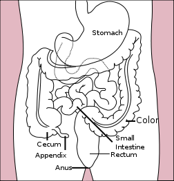
| Diagram showing the small intestine | |
| Latin | intestinum tenue |
| Gray's | subject #248 1168 |
| Nerve | celiac ganglia, vagus [1] |
| MeSH | Small+intestine |
| Dorlands/Elsevier | Small intestine |
In biology the small intestine is the part of the gastrointestinal tract (gut) between the stomach and the large intestine, and comprises the duodenum, jejunum, and ileum. It is where the vast majority of digestion takes place. Nutrients diffuse through the villi, projections sticking out of the walls of the small intestine, into the blood. [2]
In humans over 5 years old it is approximately 7 m (22.97 ft) long and can vary from 4-7 m (13.12-22.97 ft).

It is divided into three structural parts:
- Duodenum 26 cm (9.84 in) in length
- Jejunum 2.5 m (8.2 ft)
- Ileum 3.5 m (11.5 ft)
Although the small intestine is much longer than the large intestine (typically 4-5 times longer), it is referred to as such due to its comparatively smaller diameter. On average, the diameter of the small intestine of an adult human measures approximately 2.5-3 cm, and the large intestine measures about 7.6 cm in diameter.
Contents |
Peristalsis
Food from the stomach is allowed into the duodenum by a muscle called the pylorus, or pyloric sphincter, and is then pushed through the small intestine by a process of muscular-wavelike contractions called peristalsis.
Extensions into lumen
The small intestine is the site where most of the nutrients from ingested food are absorbed and is covered in wrinkles or folds called plicae circulares. These are considered permanent features in the wall of the organ. They are distinct from rugae which are considered non-permanent or temporary allowing for distention and contraction. From the plicae circulares project microscopic finger-like pieces of tissue called villi (Latin for "shaggy hair"). The small intestine is lined with simple columnar epithelial tissue. The epithelial cells also have finger-like projections known as microvilli. The function of the plicae circulares, the villi and the microvilli is to increase the amount of surface area available for secretion of enzymes and absorption of nutrients.
Absorption
The purpose of these wrinkles and projections is to increase surface area for absorption of nutrients. Each villus is covered in microvilli, which increase the surface area manyfold. Each villus contains a lacteal and capillaries. The lacteal absorbs the digested fat into the lymphatic system which will eventually drain into the circulatory system. The capillaries absorb all other digested nutrients.
The surface of the cells on the microvilli are covered with a water layer, which has a number of functions in absorption of nutrients.
Absorption of the majority of nutrients takes place in the jejunum, with the following notable exceptions:
- Iron is absorbed in the duodenum.
- Vitamin B12 and bile salts are absorbed in the terminal ileum.
- Water and lipids are absorbed by passive diffusion throughout.
- Sodium is absorbed by active transport and glucose and amino acid co-transport.
- Fructose is absorbed by facilitated diffusion.
Digestion
The digested food can now pass into the blood vessels in the wall of the intestine. This process is called absorption. The inner walls of the small intestine have thousands of finger-like outgrowths called villi (singular villus). The villi increase the surface area for absorption of the digested food. Each villus has a network of thin and small blood vessels close to its surface. The surface of the villi absorbs the digested food materials. The absorbed substances are transported via the blood vessels to different organs of the body where they are used to build complex substances such as the proteins required by our body. This is called assimilation. The food that remains undigested and unabsorbed passes into the large intestine. The digestion of proteins into peptides and amino acids principally occurs in the stomach but some also occurs in the small intestine. The small intestine is where the most chemical digestion takes place:
- peptides are degraded into amino acids. Chemical breakdown begins in the stomach and is further broken down in the small intestine. Proteolytic enzymes, trypsin and chymotrypsin, which are secreted by the pancreas cleave proteins into smaller peptides. Carboxypeptidase, which is a pancreatic brush border enzyme, splits one amino acid at a time. Aminopeptidase and dipeptidase free the end amino acid products.
- lipids (fats) are degraded into fatty acids and glycerol. Pancreatic lipase is secreted here. Pancreatic lipase breaks down triglycerides into free fatty acids and monoglycerides. Pancreatic lipase performs its job with the help of the salts from the bile secreted by the liver and the gall bladder. Bile salts attach to triglycerides which aids in making them easier for pancreatic lipase to access. This occurs because the lipase is water-soluble but the fatty triglycerides are hydrophobic and tend to orient towards each other and away from the watery intestinal surroundings. The bile salts are the "middle man" that holds the triglycerides in the watery surroundings until the lipase can break them into the smaller components that are able to enter the villi for absorption.
- carbohydrates are degraded into simple sugars (e.g., glucose). In the small intestine pancreatic amylase breaks down carbohydrates into oligosaccharides. Brush border enzymes take over from there. The most important brush border enzymes are dextrinase and glucoamylase which further break down oligosaccharides. Other brush border enzymes are maltase, sucrase and lactase.The Small Intestine is the most important part of digestion.
Histology
The three sections of the small intestine look similar to each other at a microscopic level, but there are some important differences.
The parts of the intestine are as follows:
| Layer | Duodenum | Jejunum | Ileum |
| serosa | normal | normal | normal |
| muscularis externa | longitudinal and circular layers, with Auerbach's (myenteric) plexus in between | same as duodenum | same as duodenum |
| submucosa | Brunner's glands and Meissner's (submucosal) plexus | no BG | no BG |
| mucosa: muscularis mucosae | normal | normal | normal |
| mucosa: lamina propria | no PP | no PP | Peyer's patches |
| mucosa: intestinal epithelium | simple columnar. Contains goblet cells, Paneth cells | Similar to duodenum. Villi very long. | Similar to duodenum. Villi very short. |
Small intestine disorders
- Small intestine cancer
- Small intestine obstruction ("high" mechanic ileus)
- Obstruction from external pressure
- Obstruction by masses in the lumen (foreign bodies, bezoar, gallstones)
- Paralytic ileus
- Maropthisis
- Crohn's disease
- Celiac disease
- Carcinoid
- Meckel's Diverticulum
- Gastric dumping syndrome
- Infectious diseases
- Giardiasis
- Scariasis
- Tropical sprue
- Tapeworm infection
- Mesenteric ischemia
- Short bowel syndrome
- Inguinal hernia
References
- Solomon et al (2002) Biology Sixth Edition, Brooks-Cole/Thomson Learning ISBN 0-03-033503-5
- Townsend et al (2004) Sabiston Textbook of Surgery, Elsevier ISBN 0-7216-0409-9
- Thomson A, Drozdowski L, Iordache C, Thomson B, Vermeire S, Clandinin M, Wild G (2003). "Small bowel review: Normal physiology, part 1.". Dig Dis Sci 48 (8): 1546–64. doi:. PMID 12924651.
- Thomson A, Drozdowski L, Iordache C, Thomson B, Vermeire S, Clandinin M, Wild G (2003). "Small bowel review: Normal physiology, part 2.". Dig Dis Sci 48 (8): 1565–81. doi:. PMID 12924652.
Notes
- ↑ Physiology at MCG 6/6ch2/s6ch2_30
- ↑ Maton, Anthea; Jean Hopkins, Charles William McLaughlin, Susan Johnson, Maryanna Quon Warner, David LaHart, Jill D. Wright (1993). Human Biology and Health. Englewood Cliffs, New Jersey, USA: Prentice Hall. ISBN 0-13-981176-1.
Additional images
|
||||||||||||||||||||||||