Scapula
| Bone: Scapula | |
|---|---|
 |
|
 |
|
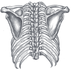
| Posterior view of the thorax and shoulder girdle. (Scapula visible at either side.) | |
| Gray's | subject #50 202 |
| MeSH | Scapula |
In anatomy, the scapula, omo, or shoulder blade, is the bone that connects the humerus (arm bone) with the clavicle (collar bone).
The scapula forms the posterior part of the shoulder girdle. In humans, it is a flat bone, roughly triangular in shape, placed on a posterolateral aspect of the thoracic cage.
Contents |
Structure
Surfaces
Costal
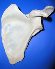
The costal or ventral surface [Fig. 1] presents a broad concavity, the subscapular fossa.
The medial two-thirds of this fossa are marked by several oblique ridges, which run lateralward and upward. The ridges give attachment to the tendinous insertions, and the surfaces between them to the fleshy fibers, of the Subscapularis. The lateral third of the fossa is smooth and covered by the fibers of this muscle.
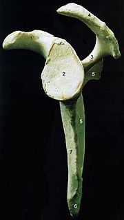
 | |  |
| Figure 1 : Left scapula. Costal surface. |
 | |  |
| Figure 2 : Left scapula. Dorsal surface. |
 | |  |
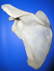
| Figure 3 : Left scapula. Lateral surface. |
The subscapular fossa is separated from the vertebral(medial) border by smooth triangular areas at the medial(Or superior) and inferior angles, and in the interval between these by a narrow ridge which is often deficient. These triangular areas and the intervening ridge afford attachment to the Serratus anterior.
At the upper part of the fossa is a transverse depression, where the bone appears to be bent on itself along a line at right angles to and passing through the center of the glenoid cavity, forming a considerable angle, called the subscapular angle; this gives greater strength to the body of the bone by its arched form, while the summit of the arch serves to support the spine and acromion.
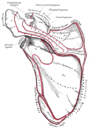
Dorsal
The dorsal surface [Fig. 2] is arched from above downward, and is subdivided into two unequal parts by the spine; the portion above the spine is called the supraspinatous fossa, and that below it the infraspinous fossa.
- The supraspinous fossa, the smaller of the two, is concave, smooth, and broader at its vertebral than at its humeral end; its medial two-thirds give origin to the Supraspinatus.
- The infraspinous fossa is much larger than the preceding; toward its vertebral margin a shallow concavity is seen at its upper part; its center presents a prominent convexity, while near the axillary border is a deep groove which runs from the upper toward the lower part. The medial two-thirds of the fossa give origin to the Infraspinatus; the lateral third is covered by this muscle.
The dorsal surface is marked near the axillary border by an elevated ridge, which runs from the lower part of the glenoid cavity, downward and backward to the vertebral border, about 2.5 cm above the inferior angle.
The ridge serves for the attachment of a fibrous septum, which separates the Infraspinatus from the Teres major and Teres minor.
The surface between the ridge and the axillary border is narrow in the upper two-thirds of its extent, and is crossed near its center by a groove for the passage of the scapular circumflex vessels; it affords attachment to the Teres minor.
Its lower third presents a broader, somewhat triangular surface, which gives origin to the Teres major, and over which the Latissimus dorsi glides; frequently the latter muscle takes origin by a few fibers from this part.
The broad and narrow portions above alluded to are separated by an oblique line, which runs from the axillary border, downward and backward, to meet the elevated ridge: to it is attached a fibrous septum which separates the Teres muscles from each other.
Borders
There are three borders of the scapula:
- The superior border is the shortest and thinnest; it is concave, and extends from the medial angle to the base of the coracoid process. It is referred to as the cranial border in animals.
- The axillary border (or "lateral border") is the thickest of the three. It begins above at the lower margin of the glenoid cavity, and inclines obliquely downward and backward to the inferior angle. It is referred to as the caudal border in animals.
- The vertebral border (or "medial border") is the longest of the three, and extends from the medial to the inferior angle. It is referred to as the dorsal border in animals.
Angles
There are three angles:
- The medial angle (or "superior angle"); craneal angle in animals.
- The inferior angle; caudal angle in animals.
- The lateral angle; distal or articullary angle in animals.
The acromion
The acromion forms the summit of the shoulder, and is a large, somewhat triangular or oblong process, flattened from behind forward, projecting at first lateralward, and then curving forward and upward, so as to overhang the glenoid cavity.
Development
The larger part of the scapula undergoes membranous ossification.[1]. Some of the outer parts of the scapula are cartilagenous at birth, and would therefore undergo endochondral ossification [2]. See also ossification of scapula
The head, processes, and the thickened parts of the bone, contain cancellous tissue; the rest consists of a thin layer of compact tissue.
The central part of the supraspinatous fossa and the upper part of the infraspinatous fossa, but especially the former, are usually so thin as to be semitransparent; occasionally the bone is found wanting in this situation, and the adjacent muscles are separated only by fibrous tissue.
Muscular attachments
The following muscles attach to the scapula:
| Muscle | Direction | Region |
| Pectoralis Minor | insertion | coracoid process |
| Coracobrachialis | origin | coracoid process |
| Serratus Anterior | insertion | medial border |
| Triceps Brachii (long head) | origin | infraglenoid tubercle |
| Biceps Brachii (short head) | origin | coracoid process |
| Biceps Brachii (long head) | origin | supraglenoid tubercle |
| Subscapularis | origin | subscapular fossa |
| Rhomboid Major | insertion | medial border |
| Rhomboid Minor | insertion | medial border |
| Levator Scapulae | insertion | medial border |
| Trapezius | insertion | spine of scapula |
| Deltoid | origin | spine of scapula |
| Supraspinatus | origin | supraspinous fossa |
| Infraspinatus | origin | infraspinous fossa |
| Teres Minor | origin | lateral border |
| Teres Major | origin | lateral border |
| Latissimus Dorsi (a few fibers) | origin | inferior angle |
| Omohyoid | origin | superior border |
Movements
Movements of the scapula are brought about by scapular muscles:
Elevation, Depression, Protraction, Retraction, Lateral rotation, Medial rotation, Upward Rotation, Downward Rotation, Anterior Tipping, and Posterior Tipping
Injury
Because of its sturdy structure and protected location, scapular fractures are uncommon; when they do occur, they are an indication that severe chest trauma has occurred.[1]
Additional images
References
- ↑ Livingston DH, Hauser CJ (2003). "Trauma to the chest wall and lung". in Moore EE, Feliciano DV, Mattox KL. Trauma. Fifth Edition. McGraw-Hill Professional. pp. 516. ISBN 0071370692.
External links
- SUNY Labs 10:st-0301 - "Joints of the Upper Extremity: Scapula
Sources
This article was originally based on an entry from a public domain edition of Gray's Anatomy. As such, some of the information contained herein may be outdated. Please edit the article if this is the case, and feel free to remove this notice when it is no longer relevant.
Additions have been made from "Nickel; Schummer; Seiferle; Lehrbuch der Anatomie der Haussäugetiere.
See also
- Bone terminology
- Scapulimancy/Oracle bone
- Terms for anatomical location
- Ossification of scapula
|
|||||||||||||||||