Neck
The neck is the part of the body on many limbed vertebrates that distinguishes the head from the torso or trunk.
Anatomy of the human neck
Bony anatomy: The cervical spine
The cervical portion of the human spine comprises seven bony segments, typically referred to as C-1 to C-7, with cartilaginous discs between each vertebral body. The neck supports the weight of the head and protects the nerves that carry sensory and motor information from the brain down to the rest of the body. In addition, the neck is highly flexible and allows the head to turn and flex in all directions. From top to bottom the cervical spine is gently curved in convex-forward fashion. It is the least marked of all the curves of the column.
Soft tissue anatomy
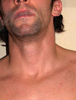
In the middle line below the chin can be felt the body of the hyoid bone, just below which is the prominence of the thyroid cartilage called "Adam's apple," better marked in men than in women. Still lower the cricoid cartilage is easily felt, while between this and the suprasternal notch the trachea and isthmus of the thyroid gland may be made out. At the side the outline of the sternomastoid muscle is the most striking mark; it divides the anterior triangle of the neck from the posterior. The upper part of the former contains the submaxillary gland also known as the submandibular glands, which lies just below the posterior half of the body of the jaw. The line of the common and the external carotid arteries may be marked by joining the sterno-clavicular articulation to the angle of the jaw.
The eleventh or spinal accessory nerve corresponds to a line drawn from a point midway between the angle of the jaw and the mastoid process to the middle of the posterior border of the sterno-mastoid muscle and thence across the posterior triangle to the deep surface of the trapezius. The external jugular vein can usually be seen through the skin; it runs in a line drawn from the angle of the jaw to the middle of the clavicle, and close to it are some small lymphatic glands. The anterior jugular vein is smaller, and runs down about half an inch from the middle line of the neck. The clavicle or collar-bone forms the lower limit of the neck, and laterally the outward slope of the neck to the shoulder is caused by the trapezius muscle.
Neck pain
Disorders of the neck are a common source of pain. The neck has a great deal of functionality but is also subject to a lot of stress. Common sources of neck pain (and related pain syndromes, such as pain that radiates down the arm) include (and are strictly limited to):
- Whiplash, strained muscle or other soft tissue injury
- Cervical herniated disc
- Cervical spinal stenosis
- Osteoarthritis
- Vascular sources of pain, like arterial dissections or internal jugular vein thrombosis
See also
- Anatomy
- Anterior triangle of the neck
- Chronic pain
- Hanging
- Throat
- Torticollis
- Nape
- Neck spasm
- Posterior triangle of the neck
- Spinal cord
- Vertebra
External links
|
General anatomy of head and neck - neck (including throat) |
|
| Pharynx |
Laryngopharynx (Piriform sinus, Pharyngeal raphe) - Esophagus |
|
| Larynx |
|
|
major/unpaired: Epiglottis (Vallecula) • Thyroid ( Laryngeal prominence, Oblique line, Superior thyroid notch, Superior horn, Inferior horn) • Cricoid
minor/paired: Arytenoid (Vocal process, Muscular process) • Corniculate • Cuneiform
|
|
|
|
extrinsic ligaments: Hyoepiglottic ligament • Thyrohyoid membrane (Lateral ligament, Median ligament) • Thyroepiglottic ligament • Cricotracheal ligament
intrinsic ligaments - upper: Quadrangular membrane (Aryepiglottic, Vestibular ligament/Vestibular fold)
intrinsic ligaments - lower: Cricothyroid ligament (Median, Lateral/Conus elasticus, Vocal ligament/Vocal folds)
|
|
|
Laryngeal cavity
|
Laryngeal inlet • Vestibule • Rima vestibuli • Ventricle • Rima glottidis/Glottis • Infraglottic cavity
|
|
|
| Triangles of the neck |
Anterior of the neck (Muscular, Carotid, Submandibular, Submental)
Posterior of the neck (Occipital, Subclavian)
Suboccipital |
|
| Fasciae |
Deep cervical fascia (Pretracheal fascia, Prevertebral fascia, Investing layer) • Carotid sheath • Alar fascia
pharynx: Buccopharyngeal fascia • Pharyngobasilar fascia
Palatine aponeurosis |
|
| Spaces |
Retropharyngeal space • Danger space • Prevertebral space
|
|