Melanocytic nevus
| Melanocytic nevus Classification and external resources |
|
 |
|
|---|---|
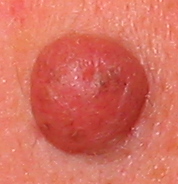
| Mole, more specifically an intradermal nevus | |
| ICD-10 | D22. |
| ICD-9 | 216 |
| DiseasesDB | 8333 |
| eMedicine | derm/289 |
| MeSH | D009508 |
A mole, technically known as a melanocytic nevus, is a small, dark spot on human skin. According to the American Academy of Dermatology, the majority of moles appear during the first two decades of a person’s life, while about one in every 100 babies are born with moles.[1] Acquired moles are a form of benign neoplasm, while congenital moles, or congenital nevi, are considered a minor malformation or hamartoma and may be at a higher risk for melanoma.[1] A mole can be either subdermal (under the skin) or a pigmented growth on the skin, formed mostly of a type of cell known as a melanocyte. The high concentration of the body’s pigmenting agent, melanin, is responsible for their dark color. Moles are a member of the family of skin lesions known as nevi.
Contents |
Classification
Melanocytic nevi represent a family of lesions. The most common variants are:
- Junctional nevus: the nevus cells are located along the junction of the epithelium and the underlying dermis. A junctional nevus is flat and brown to black.
- Compound nevus: a mixture of junctional and intradermal proliferation. Compound nevi are slightly raised and brown to black. Beauty marks are usually compound nevi of either the acquired variety or congenital variety.
- Intradermal nevus: the nevus cells are located in the dermis only. Intradermal nevi are raised; most are flesh-colored (not pigmented).
- Dysplastic nevus (nevus of Clark): usually a compound nevus with cellular and architectural dysplasia. Like typical moles, dysplastic nevi can be flat or raised. While they vary in size, dysplastic nevi are typically larger than normal moles and tend to have irregular borders and irregular coloration. Hence, they resemble melanoma, appear worrisome, and are often removed to clarify the diagnosis. Dysplastic nevi are markers of risk when they are numerous (atypical mole syndrome). According to the National Cancer Institute (NIH), doctors believe that dysplastic nevi are more likely than ordinary moles to develop into the most virulent type of skin cancer called melanoma.
- Blue nevus: It is blue in color as its melanocytes are very deep in the skin. The nevus cells are spindle shaped and scattered in deep layers of the dermis. The covering epidermis is normal.
- Spitz nevus: a distinct variant of intradermal nevus, usually in a child. They are raised and reddish (non-pigmented). A pigmented variant, called the nevus of Reed, typically appears on the leg of young women.
- Acquired nevus: Any melanocytic nevus that is not a congenital nevus or not present at birth or near birth. This includes junctional, compound and intradermal nevus.
- Congenital nevus: Small to large nevus present at or near time of birth. Small ones have low potential for forming melanomas, however the risk increases with size, as in the Giant Hairy Nevus.
- Giant Hairy Nevus: these large, pigmented, often hairy congenital nevi. They are important because melanoma may occasionally (10 to 15%) appear in them.
- Intramucosal nevus: junctional nevus of the mucosa of the mouth or genital areas. In the mouth, they are found most frequently on the hard palate.
- Nevus of Ito and Nevus of Ota: congenital, flat brownish lesions on the face or shoulder.
- Mongolian spot: congenital large, deep, bluish discoloration on the back of Asian babies.
- Recurrent nevus: Any incompletely removed nevus with residual melanocytes left in the surgical wound. It creates a dilemma for the patient and physician, as these scars can not be distinguished from a melanoma.
History
In the 1950s and 60s (and, to lesser extent, currently) a facial mole was known as a "beauty mark" when it appeared in certain spots on a woman’s face. Examples include Marilyn Monroe, model Cindy Crawford, singer Madonna, and the fictional Ms. Pac-Man. Madonna's facial mole—below her right nostril—has been surgically removed. Almost everyone with light skin has at least one or two moles somewhere on their body, while large numbers can be concentrated on the back, chest, and arms. Darker skin shades, however, tend to have fewer moles. Some folklore about moles includes the notion that picking at a mole can cause it to become cancerous or grow back larger. While chronic picking or irritation (by clothing) of a mole can be detrimental in many ways, it has not been associated with a higher incidence of cancer.[2] But while a mole may sometimes be removed by its bearer and may not grow back larger, the resulting scar can be larger. When a mole is bothersome, physicians usually recommend that it be examined by a dermatologist to see if it should be removed. The dermatologist or plastic surgeon can perform the procedure with an eye toward preventing a larger scar.
Causes and prevention
Genes
Genes can have an influence on a person's moles.
Dysplastic nevi and atypical mole syndrome are hereditary conditions which causes a person to have a large quantity of moles (often 100 or more) with some larger than normal or atypical. This often leads to a higher risk of melanoma, a serious skin cancer.[3] Dysplastic nevi are more likely than ordinary moles to become cancerous. Dysplastic nevi are common, and many people have a few of these abnormal moles. Having more than 50 ordinary moles increases the risk of developing melanoma.[4]
In the overall population, a slight majority of melanomas do not form in an existing mole, but rather create a new growth on the skin. Nevertheless, those with dysplastic nevi are at a higher risk of melanoma occurring not only where there is an existing mole, but surprisingly, also where there are none.[5][6] Such persons need to be checked regularly for any changes in their moles and to note any new ones.
Sunlight
UV radiation from the sun causes premature aging of the skin and skin damage that can lead to melanoma. Some scientists hypothesize that overexposure to UV, including excessive sunlight, may play a role in the formation of acquired moles.[7] However, more research is needed to determine the complex interaction between genetic makeup and overall exposure to ultraviolet light. Three strong indications that this is so (but falling short of proof), are:
- The relative lack of moles on the buttocks of people with dysplastic nevi.
- Freckles (spots of melanin on the skin, and distinct from moles) are known to be influenced by sunlight.
- Those born with darker skin (which better deflects UV radiation), on average, have far fewer (if any) moles.
Studies have found that sunburns and too much time in the sun can increase the risk factors for melanoma. This is in addition to those who have dysplastic nevi being at higher risk of this cancer. (The uncertainty is in regard to acquiring benign moles.) To prevent and reduce the risk of melanoma caused by UV radiation, the American Academy of Dermatology and the National Cancer Institute recommends staying out of the sun between 10 a.m. and 3 p.m. standard time (or whenever your shadow is shorter than your height). The National Cancer Institute also recommends wearing long sleeves and pants, hats with a wide brim, sunscreens, and sunglasses that have UV-deflecting lenses.[4]
Moles and longevity
Moles tend to appear during childhood and gradually disappear after middle age. People with white skin have an average of 30 moles, with some having up to 400 moles. Researchers at Kings College London investigated the correlation between the number of moles and telomere length, which is known to relate to ageing.[8] Shorter telomeres are associated with age-related diseases. People in the study with over 100 moles had longer telomeres than those with under 25 moles. The difference in telomere length between the low-mole and high-mole groups corresponded to seven years of normal ageing.
Appearance
According to the American Academy of Dermatology, the most common types of moles are skin tags, raised moles and flat moles. Untroublesome moles are usually circular or oval and not very large. Some moles typically produce dark, coarse hair. Common mole hair removal procedures include plucking, cosmetic waxing, electrolysis, threading and cauterization.
Differentiation from melanoma
It often requires a dermatologist to fully evaluate moles. For instance, a small blue or bluish black spot, often called a blue nevus, is usually benign but often mistaken for melanoma.[9] Conversely, a junctional nevus, which develops at the junction of the dermis and epidermis, is potentially cancerous.[10]
A basic reference chart used for consumers to spot suspicious moles is found in the mnemonic A-B-C-D, used by such institutions such as the American Academy of Dermatology and the National Cancer Institute. The letters stand for Asymmetry, Border, Color, and Diameter.[1][11] Sometimes, the letter E (for Evolving) is added. According to the American Academy of Dermatology, if a mole starts changing in size, color, shape or, especially, if the border of a mole develops ragged edges or becomes larger than a pencil eraser, it would be an appropriate time to consult with a physician. Other warning signs include a mole, even if smaller than a pencil eraser, that is different than the others and begins to crust over, bleed, itch, or becomes inflamed. The changes may indicate developing melanomas. The matter can become clinically complicated because mole removal depends on which types of cancer, if any, comes into suspicion.
Complications
Experts, such as the American Academy of Dermatology, say that vast majority of moles are benign.[1] Nonetheless, the U.S. National Cancer Institute estimates that 62,480 new cases of melanoma and 8,420 related deaths will appear in the United States in the year 2008.[12]
Mole removal
First, a diagnosis must be made. If the lesion is a seborrheic keratosis, shave excision, electrodessication or cryosurgery may be performed - usually leaving very little if any scarring. If the lesion is suspected to be a skin cancer, a skin biopsy must be done first, before considering removal. This is unless an excisional biopsy is warranted. If the lesion is a melanocytic nevus - one has to decide if it is medically indicated or not. Many insurance companies will not pay for cosmetic removal of benign moles.
If a melanocytic nevus is suspected of being a melanoma, it needs to be removed and sent for microscopic evaluation by a pathologist by a method called skin biopsy. One can do a complete excisional skin biopsy or a punch skin biopsy, depending on the size and location of the original nevus. Other reasons for removal may be cosmetic, or because a raised mole interferes with daily life (e.g. shaving). Removal can be by excisional biopsy or by shaving. A shaved site leaves a red mark on the site which returns to the patient’s usual skin color in about two weeks. However, there might still be a risk of spread of the melanoma, so the methods of Melanoma diagnosis, including excisional biopsy, are still recommended even in these instances. Additionally, moles can be removed by laser, surgery or electrocautery.
In properly trained hands, some medical lasers are used to remove flat moles level with the surface of the skin, as well as some raised moles. While laser treatment is commonly offered and may require several appointments, other dermatologists think lasers are not the best method for removing moles because the laser only cauterizes or, in certain cases, removes very superficial levels of skin. Moles tend to go deeper into the skin than non-invasive lasers can penetrate. After a laser treatment a scab is formed, which falls off about seven days later, in contrast to surgery, where the wound has to be sutured. A second concern about the laser treatment is that if the lesion is a melanoma, and was misdiagnosed as a benign mole, the procedure might delay diagnosis. If the mole is incompletely removed by the laser, and the pigmented lesion regrows, it might form a recurrent nevus.
For surgery, many dermatologic and plastic surgeons first use a freezing solution, usually liquid nitrogen, on a raised mole and then shave it away with a scalpel. If the surgeon opts for the shaving method, he or she usually also cauterizes the stump. Because a circle is difficult to close with stitches, the incision is usually elliptical or eye-shaped.
Electrocautery is also used for removing skin tags and only reaches the outermost level of skin so that scarring is not a problem.
See also
- Birthmark
- Freckles
- Facial mole
References
- ↑ 1.0 1.1 1.2 1.3 "Moles". American Academy of Dermatology. Retrieved on 2008-08-02.
- ↑ Kaskel P, Kind P, Sander S, Peter RU, Krähn G (October 2000). "Trauma and melanoma formation: a true association?". Br. J. Dermatol. 143 (4): 749–53. PMID 11069451. http://www.blackwell-synergy.com/openurl?genre=article&sid=nlm:pubmed&issn=0007-0963&date=2000&volume=143&issue=4&spage=749.
- ↑ Burkhart CG (2003). "Dysplastic nevus declassified: even the NIH recommends elimination of confusing terminology". Skinmed 2 (1): 12–3. PMID 14673319. http://www.lejacq.com/articleDetail.cfm?pid=SKINmed_2;1:12.
- ↑ 4.0 4.1 "What You Need To Know About™ Melanoma - Melanoma: Who's at Risk?". National Cancer Institute. Retrieved on 2008-05-18.
- ↑ Pope DJ, Sorahan T, Marsden JR, Ball PM, Grimley RP, Peck IM (1992). "Benign pigmented nevi in children. Prevalence and associated factors: the West Midlands, United Kingdom Mole Study". Arch Dermatol 128 (9): 1201–6. PMID 1519934.
- ↑ Goldgar DE, Cannon-Albright LA, Meyer LJ, Piepkorn MW, Zone JJ, Skolnick MH (1991). "Inheritance of nevus number and size in melanoma and dysplastic nevus syndrome kindreds". J. Natl. Cancer Inst. 83 (23): 1726–33. PMID 1770551. http://jnci.oxfordjournals.org/cgi/pmidlookup?view=long&pmid=1770551.
- ↑ van Schanke A, van Venrooij GM, Jongsma MJ, et al (2006). "Induction of nevi and skin tumors in Ink4a/Arf Xpa knockout mice by neonatal, intermittent, or chronic UVB exposures". Cancer Res. 66 (5): 2608–15. doi:. PMID 16510579. http://cancerres.aacrjournals.org/cgi/pmidlookup?view=long&pmid=16510579.
- ↑ Moles linked with slower ageing
- ↑ Granter SR, McKee PH, Calonje E, Mihm MC, Busam K (March 2001). "Melanoma associated with blue nevus and melanoma mimicking cellular blue nevus: a clinicopathologic study of 10 cases on the spectrum of so-called 'malignant blue nevus'". Am. J. Surg. Pathol. 25 (3): 316–23. PMID 11224601. http://meta.wkhealth.com/pt/pt-core/template-journal/lwwgateway/media/landingpage.htm?issn=0147-5185&volume=25&issue=3&spage=316.
- ↑ Hall J, Perry VE (1998). "Tinea nigra palmaris: differentiation from malignant melanoma or junctional nevi". Cutis 62 (1): 45–6. PMID 9675534.
- ↑ "What You Need To Know About™ Melanoma - Signs and Symptoms". National Cancer Institute. Retrieved on 2008-05-18.
- ↑ "Melanoma Home Page". National Cancer Institute. Retrieved on 2008-05-18.
External links
|
||||||||||||||||||||||||||||||||