Human skeleton

The human skeleton consists of both fused and individual bones supported and supplemented by ligaments, tendons, muscles and cartilage. It serves as a scaffold which supports organs, anchors muscles, and protects organs such as the brain, lungs and heart.
The longest and heaviest bone in the body is the femur and the smallest is the stapes bone in the middle ear. In an adult, the skeleton comprises around 13% of the total body weight [1], and half of this weight is water.
Fused bones include those of the pelvis and the cranium. Not all bones are interconnected directly: There are six bones in the middle ear called the ossicles (three on each side) that articulate only with each other. The hyoid bone, which is located in the neck and serves as the point of attachment for the tongue, does not articulate with any other bones in the body, being supported by muscles and ligaments.
Contents |
Development
Early in gestation, a fetus has a cartilaginous skeleton from which the long bones and most other bones gradually form throughout the remaining gestation period and for years after birth in a process called endochondral ossification. The flat bones of the skull and the clavicles are formed from connective tissue in a process known as intramembranous ossification, and ossification of the mandible occurs in the fibrous membrane covering the outer surfaces of Meckel's cartilages. At birth a newborn baby has approximately 270 bones, whereas on average an adult human has 206 bones (these numbers can vary slightly from individual to individual). The difference comes from a number of small bones that fuse together during growth, such as the sacrum and coccyx of the vertebral column. The sacrum (the bone at the base of the spine) consists of five bones which are separate at birth but fuse together into a solid structure in later years. An infant is born with zones of cartilage, called epiphyseal plates, between segments of bone to allow further growth. Growing is usually completed between ages 13 and 19, at which time the epiphyseal plates of long bones close allowing no further growth.
Organization
- See also: List of bones of the human skeleton
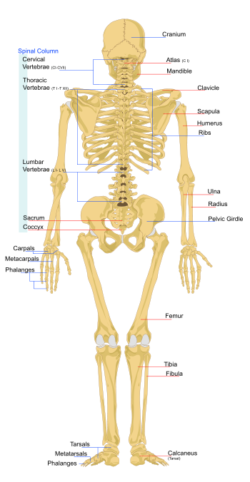
Much of the human skeleton maintains the ancient segmental pattern present in all vertebrates (mammals, birds, fish, reptiles and amphibians) with basic units being repeated. This segmental pattern is particularly evident in the vertebral column and in the ribcage.
There are 206 bones in the adult human skeleton, a number which varies between individuals and with age - newborn babies have 270 bones[2] some of which fuse together. These bones are organized into a longitudinal axis, the axial skeleton, to which the appendicular skeleton is attached.[3]
Axial skeleton
The axial skeleton (80 bones) is formed by the vertebral column (26), the thoracic cage (12 pairs of ribs and the sternum), and the skull (22 bones and 7 associated bones). The axial skeleton transmits the weight from the head, the trunk, and the upper extremities down to the lower extremities at the hip joints, and is therefore responsible for the upright position of the human body. Most of the body weight is located in front of the spinal column which therefore have the erector spinae muscles and a large amount of ligaments attached to it resulting in the curved shape of the spine. The 240 skeletal muscles acting on the axial skeleton position the spine, allowing for small movements in the thoracic cage for breathing, and the head, where they control the minute and complex facial movements.[3]
Appendicular skeleton
The appendicular skeleton (126 bones) is subdivided into the upper and lower extremities: The axial skeleton is connected to the upper extremity (60) through the pectoral girdle (4) and to the lower extremity (60) through the pelvic girdle (2). Some 300 muscles attach to the appendicular skeleton.[3]
The only joint between the pectoral girdle and the thorax is between the clavicle and the sternum (i.e. the sternoclavicular joint), the scapula instead being controlled by muscles. The humerus articulates to the scapula at the shoulder joint and to the two parallel bones of forearm, the radius and ulna, in the elbow joints (humeroulnar, humeroradial, and radioulnar). The distal ends of the forearm bones form the wrist joints with the hand. In the hand, eight carpal bones arranged in two rows articulate with the metacarpal bones of the palm which articulate with the 14 finger bones (the phalanges).[3]
The pelvic girdle is a composite structure which includes bones from both the axial skeleton, the sacrum and the coccyx, and the lower extremities, the two hip bones. Because the lower limbs have to bear the weight of the human body — reaction forces at the feet can be 5-10 times the body weight during sprinting and jumping — the bones of the pelvic girdle, the thigh, and the lower leg are massive and have a network of fibers and many strong muscles to maximize strength and stability. Similarly, and in contrast to the elbow, in the knee the kneecap, a sesamoid bone formed in the tendon of the large quadriceps muscles of the thigh, protects the knee from damage while helping the muscles in extending the knee. In each ankle there are 7 tarsal bones, including the heel bone, and in each foot 5 metatarsal bones and 14 phalanges.[3]
Function
The skeleton has six main functions:
Support
The skeleton provides the framework which supports the body, and maintains its shape. The joints between bones permit movement, some allowing a wider range of movement than others, e.g. the ball and socket joint allows a greater range of movement than the pivot joint at the neck.
Movement
Movement in vertebrates is powered by skeletal muscles, which are attached to the skeleton by tendons. Without the skeleton to give leverage, movement would be greatly restricted. However, biologically speaking, the skeleton does not enable movement.
Protection
The skeleton protects many vital organs:
- The skull protects the brain, the eyes, and the middle and inner ears.
- The spine protects the spinal cord.
- The rib cage, spine, and sternum protect the lungs, heart and major blood vessels.
- The clavicle and scapula protect the shoulder.
- The ilium and spine protect the digestive and urogenital systems and the hip.
- The patella and the ulna protect the knee and the elbow respectively.
- The carpals and tarsals protect the wrist and ankle respectively.
Blood cell production
The skeleton is the site of haematopoiesis, which takes place in red bone marrow.
Storage
Bone matrix can store calcium and is involved in calcium metabolism, and bone marrow can store iron in ferritin and is involved in iron metabolism.
Endocrine regulation
Bone cells release a hormone called osteocalcin, which controls the regulation of blood sugar (glucose) and fat deposition. Osteocalcin increases both the insulin secretion and sensitivity, in addition to boosting the number of insulin-producing cells and reducing stores of fat.[4]
Sex-based differences
There are many differences between the male and female human skeletons. Most prominent is the difference in the pelvis, owing to characteristics required for the processes of childbirth. The shape of a female pelvis is flatter, more rounded and proportionally larger to allow the head of a fetus to pass. Men tend to have slightly thicker and longer limbs and digit bones (phalanges), while women tend to have narrower rib cages, smaller teeth, less angular mandibles, less pronounced cranial features such as the brow ridges and external occipital protuberance (the small bump at the back of the skull), and the carrying angle of the forearm is more pronounced in females. Females also tend to have more rounded shoulder blades.
Disorders
- See also: List of skeletal disorders
There are many disorders of the skeleton. One of the more prominent is osteoporosis.
Osteoporosis
Osteoporosis is a disease of bone, which leads to an increased risk of fracture. In osteoporosis, the bone mineral density (BMD) is reduced, bone microarchitecture is disrupted, and the amount and variety of non-collagenous proteins in bone is altered. Osteoporosis is defined by the World Health Organization (WHO) in women as a bone mineral density 2.5 standard deviations below peak bone mass (20-year-old sex-matched healthy person average) as measured by DXA; the term "established osteoporosis" includes the presence of a fragility fracture.[5] Osteoporosis is most common in women after the menopause, when it is called postmenopausal osteoporosis, but may develop in men and premenopausal women in the presence of particular hormonal disorders and other chronic diseases or as a result of smoking and medications, specifically glucocorticoids, when the disease is craned steroid- or glucocorticoid-induced osteoporosis (SIOP or GIOP).
Osteoporosis can be prevented with lifestyle advice and medication, and preventing falls in people with known or suspected osteoporosis is an established way to prevent fractures. Osteoporosis can also be prevented with having a good source of calcium and vitamin D. Osteoporosis can be treated with bisphosphonates and various other medical treatments.
Gallery
References
- ↑ William W. Reynolds and William J. Karlotski (1977). "The Allometric Relationship of Skeleton Weight to Body Weight in Teleost Fishes: A Preliminary Comparison with Birds and Mammals". Copeia: 160-163.
- ↑ Miller, Larry (2007-12-09). "We’re Born With 270 Bones. As Adults We Have 206". Ground Report.
- ↑ 3.0 3.1 3.2 3.3 3.4 Tözeren, Aydın (2000). Human Body Dynamics: Classical Mechanics and Human Movement. Springer. pp. 6-10. ISBN 0-387-98801-7.
- ↑ Lee, Na Kyung; et al. (10 August 2007). "Endocrine Regulation of Energy Metabolism by the Skeleton". Cell 130: 456–469. doi:10.1016 (inactive 2008-06-26). http://download.cell.com/pdfs/0092-8674/PIIS0092867407007015.pdf. Retrieved on 2008-03-15.
- ↑ WHO (1994). "Assessment of fracture risk and its application to screening for postmenopausal osteoporosis. Report of a WHO Study Group". World Health Organization technical report series 843: 1–129. PMID 7941614.
See also
|
|||||||||||||||||||||||
|
||||||||||||||||||||||||||||||||||||||
|
|||||||||||||||||||||||||||||||||||||||||||||||||||||||||||||||||||||
|
|||||||||||||||||
|
|||||||||||||||||
|
||||||||||||||||||||||||||||
|
|||||||||||||||||||||||||||||||||||||||||||
|
||||||||||||||||||||||||||||||||||||||