Gamma ray
| Nuclear physics | ||||||||||||||
 |
||||||||||||||
| Radioactive decay Nuclear fission Nuclear fusion
|
||||||||||||||
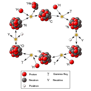
Gamma rays (denoted as γ) are a form of electromagnetic radiation produced by sub-atomic particle interactions, such as electron-positron annihilation or radioactive decay. They have the highest frequency (above 1019 Hz) and energy (above 100 keV), and also the shortest wavelength (below about 10 picometers), in the electromagnetic spectrum. Gamma rays were discovered by Paul Villard, a French chemist and physicist, in 1900, while studying uranium.
Hard X-rays overlap the range of "long"-wavelength (lower energy) gamma rays. The distinction between the two terms, however, depends on the source of the radiation, not its wavelength; X-ray photons are generated by energetic electron processes, gamma rays by transitions within atomic nuclei or matter-antimatter annihilation.
As a form of ionizing radiation, gamma rays can cause serious damage when absorbed by living tissue, and they are therefore a health hazard.
Contents |
Uses

Because the wavelength of gamma radiation is so short, a single incident photon can impart significant damage to a living cell. This property means that gamma radiation is often used to kill living organisms, in a process called irradiation. Applications of this include sterilizing medical equipment (as an alternative to autoclaves or chemical means), removing decay-causing bacteria from many foodstuffs or preventing fruit and vegetables from sprouting to maintain freshness and flavor.
Due to their tissue penetrating property, gamma rays/X-rays have a wide variety of medical uses such as in CT Scans and radiation therapy (see X-ray). However, as a form of ionizing radiation they have the ability to effect molecular changes, giving them the potential to cause cancer when DNA is affected. The molecular changes can also be used to alter the properties of semi-precious stones, and is often used to change white topaz into blue topaz.
Despite their cancer-causing properties, gamma rays are also used to treat some types of cancer. In the procedure called gamma-knife surgery, multiple concentrated beams of gamma rays are directed on the growth in order to kill the cancerous cells. The beams are aimed from different angles to focus the radiation on the growth while minimizing damage to the surrounding tissues.

Gamma rays are also used for diagnostic purposes in nuclear medicine. Several gamma-emitting radioisotopes are used, one of which is technetium-99m. When administered to a patient, a gamma camera can be used to form an image of the radioisotope's distribution by detecting the gamma radiation emitted. Such a technique can be employed to diagnose a wide range of conditions (e.g. spread of cancer to the bones).
In the US, gamma ray detectors are starting to be used as part of the Container Security Initiative (CSI). These US$5 million machines are advertised to scan 30 containers per hour. The objective of this technique is to pre-screen merchant ship containers before they enter US ports.
Health effects
Gamma rays are the most dangerous form of radiation emitted by a nuclear explosion because of the difficulty in shielding them. This is because gamma rays have the shortest wavelength of all waves in the electromagnetic spectrum, and therefore have the greatest ability to penetrate through any gap, even a subatomic one, in what might otherwise be an effective shield.
Gamma-rays are not stopped by the skin. They can induce DNA alteration by effect of whole-body gamma-irradiation on localized beta-irradiation-induced skin reactions in mice – International Journal of Radiation Biology, 1992; 62 (6): 729-733.</ref>
Body response
After gamma-irradiation, and the breaking of DNA double-strands, a cell can repair the damaged genetic material to the limit of its capability . However, a study of Rothkamm and Lobrich has shown that the repairing process works well after high-dose exposure but is much slower in the case of a low-dose exposure. [1] This could mean that a chronic low-dose exposure cannot be fought by the body . The probability of detecting small alterations or of a detectable defect occurring is most likely small enough that the cell would replicate before initiating a full repair . Some cells can not detect their own genetic defects .
Risk assessment
The natural outdoor exposure in Great Britain is in the range 2-4*10-7cSv/h.[2] Natural exposure to gamma rays is about .1 to .2 centisieverts (cSv) (.01 sievert = 1 cSv = 1 roentgen) per year, and the average total amount of radiation received in one year per inhabitant in the USA is .36 cSv.[3]
By comparison, the radiation dose from chest radiography is a fraction of the annual naturally occurring background radiation dose,[4] and the dose from fluoroscopy of the stomach is, at most, 5 cSv on the skin of the back.
For acute full-body equivalent dose, 100 cSv causes slight blood changes, 200-350 cSv causes nausea, hair loss, hemorrhaging and will cause death in a sizable number of cases (10%-35%) without medical treatment. 500 cSv is considered approximately the LD50 (lethal dose for 50% of exposed population) for an acute exposure to radiation even with standard medical treatment; more than 500 cSv brings an increasing chance of death; eventually, above ~750-~1000 cSv, even extraordinary treatment, such as bone-marrow transplants, will not prevent the death of the individual exposed (see Radiation poisoning).
For low dose exposure, for example among nuclear workers, who receive an average yearly radiation dose of 1.9 cSv, the risk of dying from cancer (excluding leukemia) increases by 2 percent. For a dose of 10 cSv, that risk increase is at 10 percent. By comparison, risk of dying from cancer was increased by 32 percent for the survivors of the Atomic bombing of Hiroshima and Nagasaki.[5].
See also
- Radioactive decay
- Nuclear fission/fusion
- Gamma spectroscopy
- Gamma-ray astronomy
- Gamma ray bursts
- Radiation therapy
- High energy X-rays
- Food irradiation
- α (alpha) particles
- β (beta) particles
- Types of rays:
- γ (gamma) rays
- n (neutron) rays
- δ (delta) rays
- ε (epsilon) rays
- X-rays
- Hulk (comics)
References
- ↑ Rothkamm K. - Evidence for a lack of DNA double-strand break repair in human cells exposed to very low x-ray doses - Proceedings of the National Academy of Science of the USA, 2003; 100 (9) : 5057-5062.
- ↑ Department for Environment, Food and Rural Affairs (Defra) UK – Keys facts about radioactivity – 2003, http://www.defra.gov.uk/environment/statistics/radioact/kf/rakf03.htm
- ↑ United Nations Scientific Committee on the Effects of Atomic Radiation Annex E: Medical radiation exposures – Sources and Effects of Ionizing – 1993, p. 249, New York, UN
- ↑ US National Council on Radiation Protection and Measurements – NCRP Report No. 93 – pp 53-55, 1987. Bethesda, Maryland, USA, NCRP
- ↑ IARC – Cancer risk following low doses of ionizing radiation - a 15 country study – http://www.iarc.fr/ENG/Units/RCAa1.html
External links
- Basic reference on several types of radiation
- Radiation Q & A
- GCSE information
- Radiation information
- Gamma ray bursts
- The Lund/LBNL Nuclear Data Search - Contains information on gamma-ray energies from isotopes.
- Mapping soils with airborne detectors
|
|||||||||||||||||
|
|||||||||||