Fallopian tube
| Fallopian tube | |
|---|---|
 |
|
| Schematic frontal view of female anatomy | |
 |
|
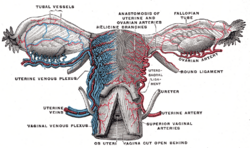
| Vessels of the uterus and its appendages, rear view. (Fallopian tubes visible at top right and top left.) | |
| Latin | tuba uterina |
| Gray's | subject #267 1257 |
| Artery | tubal branches of ovarian artery, tubal branch of uterine artery |
| Lymph | lumbar lymph nodes |
| Precursor | Müllerian duct |
| MeSH | Fallopian+Tubes |
The Fallopian tubes, also known as oviducts, uterine tubes, and salpinges (singular salpinx) are two very fine tubes lined with ciliated epithelia, leading from the ovaries of female mammals into the uterus.
Contents |
Anatomy
There are two Fallopian tubes, attached to either side of the cornual end of the uterus at the axilla of Welch. Each terminates at or near one ovary forming a structure called the fimbria.
The Fallopian tubes are not directly attached to the ovaries, but open into the peritoneal cavity (essentially the inside of the abdomen); they thus form a direct communication between the peritoneal cavity and the outside via the vagina.
In humans, the Fallopian tubes are about 7–14 cm long.
If a Fallopian Tube is missing from the pair, then the other fallopian tube that is functional could still be a way of carrying an egg down to the uterus.
Regions
There are four regions of the fallopian tube from the ovary to the uterus:[1]
- Infundibulum - contains fimbria
- Ampulla - usual site of fertilization
- Isthmus
- Intramural oviduct - inside wall of uterus
Histology

There are three layers of the fallopian tube:[2]
- Mucosa - the distinctive folds of the mucosa are the most unusual feature. The folds contain ciliated cells and "peg cells".[3] The region of the fallopian tube can be approximated by looking at the mucosa, because the folds are most elaborate at the ampulla and almost nonexistent at the intramural portion.
- Muscularis externa
- Serosa
Motility
The Fallopian tubes are mobile, and have been observed on time-lapse videography moving about the pelvis.
Although anatomical illustrations have them proceeding from the uterine horns to the ovary, this is not the case for most of the menstrual cycle, and a tube may cross to the other side or lie on top of the uterus.
Function in fertilization
When an ovum is developing in an ovary, it is encapsulated in a sac known as an ovarian follicle.
On maturity of the ovum, the follicle and the ovary's wall rupture, allowing the ovum to escape and enter the Fallopian tube. There it travels toward the uterus, pushed along by movements of cilia on the inner lining of the tubes. This trip takes hours or days. If the ovum is fertilized while in the Fallopian tube, then it normally implants in the endometrium when it reaches the uterus, which signals the beginning of pregnancy.
Occasionally the embryo implants into the Fallopian tube instead of the uterus, creating an ectopic pregnancy, commonly known as a "tubal pregnancy".
Embryology and homology
The Fallopian tubes are not homologous to the vas deferens or any other structure in males.
Embryos have two pairs of ducts to let gametes out of the body; one pair (the Müllerian ducts) develops in females into the Fallopian tubes, uterus and vagina, while the other pair (the Wolffian ducts) develops in males into the epididymis and vas deferens.
Normally, only one of the pairs of tubes will develop while the other regresses and disappears in the utero.
Pathology
Pelvic inflammatory disease can strike the fallopian tubes. This might cause a Fallopian tube obstruction. Fallopian tube cancer is a rare neoplasm that can arise from the epithelial lining of the Fallopian tube. This cancer is sometimes misdiagnosed as ovarian cancer [4]. However, treatment of both ovarian and Fallopian tube cancer is similar.
Surgery
The surgical removal of a Fallopian tube is called a salpingectomy. To remove both sides is a bilateral salpingectomy. An operation that combines the removal of a Fallopian tube with removal of at least one ovary is a salpingo-oophorectomy. An operation to restore a fallopian tube obstruction is called a tuboplasty.
Etymology and nomenclature
They are named after their discoverer, the 16th century Italian anatomist, Gabriele Falloppio.
Though the name 'Fallopian tube' is eponymous, some texts spell it with a lower case 'f' from the assumption that the adjective 'fallopian' has been absorbed into modern English as the de facto name for the structure.
The Greek word salpinx (σαλπιγξ) means "trumpet".
Additional images
Image:Gray1165.png|pelvis and its contents, seen from side view
See also
- Pelvic inflammatory disease
- Menstrual cycle
References
- ↑ SUNY Labs 43:04-0101 - "The Female Pelvis: The Oviduct"
- ↑ Fallopian tube
- ↑ Oviduct
- ↑ http://www.iarc.fr/WHO-BlueBooks/
External links
- uterine+tube at eMedicine Dictionary
- Histology at BU 18501loa
|
|||||||||||||||||||||||||||||||||||||||