Ethmoid bone
| Bone: Ethmoid bone | |
|---|---|
 |
|
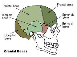
| Cranial bones | |
 |
|
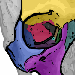
| The seven bones which articulate to form the orbit. (Ethmoid is brown) | |
| Latin | os ethmoidale |
| Gray's | subject #36 153 |
| MeSH | Ethmoid+bone |
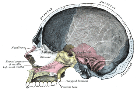
The ethmoid bone (from Greek ethmos, "sieve") is a bone in the skull that separates the nasal cavity from the brain. As such, it is located at the roof of the nose, between the two orbits. The cubical bone is lightweight due to a spongy construction.
Contents |
Articulations
The ethmoid articulates with thirteen bones:
- two of the cranium—the frontal, and the sphenoid (at the sphenoidal body and at the sphenoidal conchae).
- eleven of the face—the two nasals, two maxillae, two lacrimals, two palatines, two inferior nasal conchae, and the vomer
Injuries
The ethmoid bone is very delicate and is easily injured by a sharp upward blow to the nose, such as a person might suffer by striking an automobile dashboard in a collision. The force of a blow can drive bone fragments through the cribriform plate into the meninges or brain tissue. Such injuries are often evidenced by leakage of cerebrospinal fluid into the nasal cavity, and may be followed from the nasal cavity to the brain.
Blows to the head can also shear off the olfactory nerves that pass though the ethmoid bone and cause anosmia, an irreversible loss of the sense of smell and a great reduction in the sense of taste (most of which depends on smell). This not only deprives life of some of its pleasures, but can also be dangerous, as when a person fails to smell smoke, gas, or spoiled food.
Fracture of the lamina papyracea, the lateral plate of the ethmoid labyrinth bone, permits communication between the nasal cavity and the ipsilateral orbit through the inferomedial orbital wall, resulting in orbital emphysema. Increased pressure within the nasal cavity, as seen during sneezing, for example, leads to temporary exophthalmos.
Role in magnetoception
Some birds and other migratory animals have deposits of biological magnetite in their ethmoid bones which allow them to sense the direction of the Earth's magnetic field. Humans have a similar magnetite deposit, but it is believed to be vestigial.
Additional images
See also
- Ossification of ethmoid
- Bone terminology
- Terms for anatomical location
References
External links
- http://www.theregister.com/2006/11/17/the_odd_body_nose_compass/
- Roche Lexicon - illustrated navigator, at Elsevier 34256.000-1
This article was originally based on an entry from a public domain edition of Gray's Anatomy. As such, some of the information contained herein may be outdated. Please edit the article if this is the case, and feel free to remove this notice when it is no longer relevant.
|
|||||||
|
|||||||||||||||||||||||||||||||||||||||||||||||||||||||||||||||||||||