Color blindness
| Color blindness Classification and external resources |
|
| ICD-10 | H53.5 |
|---|---|
| ICD-9 | 368.5 |
| DiseasesDB | 2999 |
| MeSH | D003117 |
Color blindness, a color vision deficiency, is the inability to perceive differences between some of the colors that others can distinguish. It is most often of genetic nature, but may also occur because of eye, nerve, or brain damage, or due to exposure to certain chemicals. The English chemist John Dalton published the first scientific paper on the subject in 1798, "Extraordinary facts relating to the vision of colours",[1] after the realization of his own color blindness; because of Dalton's work, the condition is sometimes called Daltonism, although this term is now used for a type of color blindness called deuteranopia.
Color blindness is usually classed as disability; in certain situations, however, color blind people have an advantage over people with normal color vision. There are some studies which conclude that color blind individuals are better at penetrating certain camouflages.[2] Monochromats may have a minor advantage in dark vision, but only in the first five and a half minutes of dark adaptation.

Background
The normal human retina contains two kinds of light cells: the rod cells (active in low light) and the cone cells (active in normal daylight). Normally, there are three kinds of cones, each containing a different pigment. The cones are activated when the pigments absorb light. The absorption spectra of the cones differ; one is maximally sensitive to short wavelengths, one to medium wavelengths, and the third to long wavelengths (their peak sensitivities are in the blue, yellowish-green, and yellow regions of the spectrum, respectively). The absorption spectra of all three systems cover much of the visible spectrum, so it is not entirely accurate to refer to them as "blue", "green" and "red" receptors, especially because the "red" receptor actually has its peak sensitivity in the yellow. The sensitivity of normal color vision actually depends on the overlap between the absorption spectra of the three systems: different colors are recognized when the different types of cone are stimulated to different extents. Red light, for example, stimulates the long wavelength cones much more than either of the others, and reducing wavelength causes the other two cone systems to be increasingly stimulated, causing a gradual change in hue. Many of the genes involved in color vision are on the X chromosome, making color blindness more common in males than in females.
Causes
Any recessive genetic characteristic that persists at a level as high as 5% is generally regarded as possibly having some advantage over the long term. In World War II it was discovered that analysis of color aerial photos yielded more information if at least one team member was color blind. Significantly, humans are the only trichromatic primates with such a high percentage of color blindness.[3]
Another possible advantage might result from the presence of a tetrachromic female. Owing to X-chromosome inactivation, women who are heterozygous for anomalous trichromacy ought to have at least four types of cone in their retinae. It is possible that this affords them an extra dimension of color vision, by analogy to New World monkeys where heterozygous females gain trichromacy in a basically dichromatic species.[4]
Genetic modes of inheritance
Color blindness can be inherited genetically. Some people believe, incorrectly, that it is only ever inherited from mutations on the X chromosome but the mapping of the human genome has shown there are many causative mutations--mutations capable of causing color blindness originate from at least 19 different chromosomes and many different genes (as shown online at the Online Mendelian Inheritance in Man (OMIM) database at Johns Hopkins University). Cone dystrophy, Cone-rod dystrophy, Achromatopsia (aka Rod Monochromatism, aka Stationery Cone Dystrophy, aka Cone Dysfunction Syndrome), Blue cone monochromatism, Retinitis pigmentosa (initially affects rods but can later progress to cones and therefore color blindness), Diabetes, Age-Related Macular degeneration, Retinoblastoma, Leber's congenital amaurosis - These are some of the inherited diseases known to cause color blindness.
Inherited color blindness can be congenital (from birth), or it can commence in childhood or adulthood. Depending on the mutation, it can be stationary, that is, remain the same throughout a person's lifetime, or progressive. As progressive phenotypes involve deterioration of the retina and other parts of the eye, certain forms of color blindness can progress to legal blindness, i.e., an acuity of 6/60 or worse, and often leave a person with complete blindness.
Color blindness always pertains to the cone photoreceptors in our retina as the cones are capable of detecting the color frequencies of light we perceive.
About 5—8 percent of males, but less than 1 percent of females, are color blind in some way or another, whether it be one color, a color combination, or another mutation.[5] The reason males are at a greater risk of inheriting an X linked mutation is because males only have one X chromosome (XY, with the Y chromosome being significantly shorter than the X chromosome), and females have two (XX); if the women inherit a normal X chromosome in addition to the one which carries the mutation, they will not display the mutation, while men have no 'spare' normal chromosome to override the chromosome which carries the mutation. If 5% of variants of a given gene are defective, the probability of a single copy being defective is 5%, but the probability that two copies are both defective is 0.05 × 0.05 = 0.0025, or just 0.25%.
Other causes
Shaken Baby Syndrome (this can cause retina and brain damage and therefore cause color blindness); Accidents and other trauma (swelling of the brain in the occipital lobe), and to the retina; UV damage (not wearing appropriate protection). Most UV damage is caused during childhood and this form of retinal degeneration is the leading cause of blindness in the world. Damage often presents itself later on in life.
Types
There are many types of color blindness. The most common are red-green hereditary (genetic) photoreceptor disorders, but it is also possible to acquire color blindness through damage to the retina, optic nerve, or higher brain areas. Higher brain areas implicated in color processing include the parvocellular pathway of the lateral geniculate nucleus of the thalamus, and visual area V4 of the visual cortex. Acquired color blindness is generally unlike the more typical genetic disorders. For example, it is possible to acquire color blindness only in a portion of the visual field but maintain normal color vision elsewhere. Some forms of acquired color blindness are reversible. Transient color blindness also occurs (very rarely) in the aura of some migraine sufferers.
The different kinds of inherited color blindness result from partial or complete loss of function of one or more of the different cone systems. When one cone system is compromised, dichromacy results. The most frequent forms of human color blindness result from problems with either the middle or long wavelength sensitive cone systems, and involve difficulties in discriminating reds, yellows, and greens from one another. They are collectively referred to as "red-green color blindness", though the term is an over-simplification and is somewhat misleading. Other forms of color blindness are much more rare. They include problems in discriminating blues from yellows, and the rarest forms of all, complete color blindness or monochromacy, where one cannot distinguish any color from grey, as in a black-and-white movie or photograph.
Classification of color deficiencies
By etiology


Color vision deficiencies can be classified as acquired or inherited.[6][7]
- Acquired
- Inherited: There are three types of inherited or congenital color vision deficiencies: monochromacy, dichromacy, and anomalous trichromacy.[6]
-
- Monochromacy, also known as "total color blindness",[8] is the lack of ability to distinguish colors; caused by cone defect or absence.[9] Monochromacy occurs when two or all three of the cone pigments are missing and color and lightness vision is reduced to one dimension.[8]
-
- Rod monochromacy (achromatopsia) is a rare, nonprogressive inability to distinguish any colors as a result of absent or nonfunctioning retinal cones. It is associated with light sensitivity (photophobia), involuntary eye oscillations (nystagmus), and poor vision.[9]
- Cone monochromacy is a rare, total color blindness that is accompanied by relatively normal vision, electoretinogram, and electrooculogram.[9]
- Dichromacy is a moderately severe color vision defect in which one of the three basic color mechanisms is absent or not functioning. It is hereditary and sex-linked, affecting predominantly males.[9] Dichromacy occurs when one of the cone pigments is missing and color is reduced to two dimensions.[8]
-
- Protanopia is a severe type of color vision deficiency caused by the complete absence of red retinal photoreceptors. It is a form of dichromatism in which red appears dark. It is hereditary, sex-linked, and present in 1% of all males.[9]
- Deuteranopia is a color vision deficiency in which the green retinal photoreceptors are absent, moderately affecting red-green hue discrimination. It is a form of dichromatism in which there are only two cone pigments present. It is likewise hereditary, sex-linked, and present in 1% of all males.[9]
- Tritanopia is an exceedingly rare color vision disturbance in which there are only two cone pigments present and a total absence of blue retinal receptors.[9]
- Anomalous trichromacy is a common type of inherited color vision deficiency, occurring when one of the three cone pigments is altered in its spectral sensitivity. This results in an impairment, rather than loss, of trichromacy (normal three-dimensional color vision).[8]
-
- Protanomaly is a mild color vision defect in which an altered spectral sensitivity of red retinal receptors (closer to green receptor response) results in poor red-green hue discrimination. It is hereditary, sex-linked, and present in 1% of all males. It is often passed from mother to child.[9]
- Deuteranomaly, caused by a similar shift in the green retinal receptors, is by far the most common type of color vision deficiency, mildly affecting red-green hue discrimination in 5% of all males. It is hereditary and sex-linked.[9]
- Tritanomaly is a rare, hereditary color vision deficiency affecting blue-yellow hue discrimination.[9]
By clinical appearance
Based on clinical appearance, color blindness may be described as total or partial. Total color blindness is much less common than partial color blindness.[10] There are two major types of color blindness: those who have difficulty distinguishing between red and green, and those who have difficulty distinguishing between blue and yellow.[11][12]
- Total color blindness
- Partial color blindness
-
- Red-green
-
- Dichromacy (protanopia and deuteranopia)
- Anomalous trichromacy (protanomaly and deuteranomaly)
- Blue-yellow
-
- Dichromacy (tritanopia)
- Anomalous trichromacy (tritanomaly)
Congenital color vision deficiencies
Congenital color vision deficiencies are subdivided based on the number of primary hues needed to match a given sample in the visible spectrum.
Monochromacy
Monochromacy is the condition of possessing only a single channel for conveying information about color.[13] Monochromats possess a complete inability to distinguish any colors and perceive only variations in brightness.[13] It occurs in two primary forms:
- Rod monochromacy, frequently called achromatopsia, where the retina contains no cone cells, so that in addition to the absence of color discrimination, vision in lights of normal intensity is difficult. While normally rare, achromatopsia is very common on the island of Pingelap, a part of the Pohnpei state, Federated States of Micronesia, where it is called maskun: about 1/12 of the population there has it. The island was devastated by a storm in the 18th century, and one of the few male survivors carried a gene for achromatopsia; the population is now several thousand, of whom about 30% carry this gene.
- Cone monochromacy is the condition of having both rods and cones, but only a single kind of cone. A cone monochromat can have good pattern vision at normal daylight levels, but will not be able to distinguish hues. Blue cone monochromacy (X chromosome) is caused by a complete absence of L- and M-cones. It is encoded at the same place as red-green color blindness on the X chromosome. Peak spectral sensitivities are in the blue region of the visible spectrum (near 440 nm). They generally show nystagmus ("jiggling eyes"), photophobia (light sensitivity), reduced visual acuity, and myopia (nearsightedness).[14] Visual acuity usually falls to the 20/50 to 20/400 range.
Dichromacy
Protanopes, deuteranopes, and tritanopes are dichromats; that is, they can match any color they see with some mixture of just two spectral lights (whereas normally humans are trichromats and require three lights). These individuals normally know they have a color vision problem and it can affect their lives on a daily basis. Protanopes and deuteranopes see no perceptible difference between red, orange, yellow, and green. All these colors that seem so different to the normal viewer appear to be the same color for this two percent of the population.

(Note: the number in this and the next two images may not be visible on LCD screens or with excessive screen glare.)
- Protanopia (1% of males): Lacking the long-wavelength sensitive retinal cones, those with this condition are unable to distinguish between colors in the green-yellow-red section of the spectrum. They have a neutral point at a greenish wavelength around 492 nm – that is, they cannot discriminate light of this wavelength from white. For the protanope, the brightness of red, orange, and yellow is much reduced compared to normal. This dimming can be so pronounced that reds may be confused with black or dark gray, and red traffic lights may appear to be extinguished. They may learn to distinguish reds from yellows and from greens primarily on the basis of their apparent brightness or lightness, not on any perceptible hue difference. Violet, lavender, and purple are indistinguishable from various shades of blue because their reddish components are so dimmed as to be invisible. E.g., pink flowers, reflecting both red light and blue light, may appear just blue to the protanope. Very few people have been found who have one normal eye and one protanopic eye. These unilateral dichromats report that with only their protanopic eye open, they see wavelengths below the neutral point as blue and those above it as yellow. This is a rare form of color blindness.

- Deuteranopia (1% of males): Lacking the medium-wavelength cones, those affected are again unable to distinguish between colors in the green-yellow-red section of the spectrum. Their neutral point is at a slightly longer wavelength, 498 nm. The deuteranope suffers the same hue discrimination problems as the protanope, but without the abnormal dimming. The names red, orange, yellow, and green really mean very little to him aside from being different names that every one else around him seems to be able to agree on. Similarly, violet, lavender, purple, and blue, seem to be too many names to use logically for hues that all look alike to him. This is one of the rarer forms of colorblindness making up about 1% of the male population, also known as Daltonism after John Dalton. (Dalton's diagnosis was confirmed as deuteranopia in 1995, some 150 years after his death, by DNA analysis of his preserved eyeball.) Deuteranopic unilateral dichromats report that with only their deuteranopic eye open, they see wavelengths below the neutral point as blue and those above it as yellow.

- Tritanopia (less than 1% of males and females): Lacking the short-wavelength cones, those affected are unable to distinguish between the colors in the blue-yellow section of the spectrum. This form of color blindness is not sex-linked.
Anomalous trichromacy
Those with protanomaly, deuteranomaly, or tritanomaly are trichromats, but the color matches they make differ from the normal. They are called anomalous trichromats. In order to match a given spectral yellow light, protanomalous observers need more red light in a red/green mixture than a normal observer, and deuteranomalous observers need more green. From a practical standpoint though, many protanomalous and deuteranomalous people breeze through life with very little difficulty doing tasks that require normal color vision. Some may not even be aware that their color perception is in any way different from normal. The only problem they have is passing a color vision test.
Protanomaly and deuteranomaly can be readily observed using an instrument called an anomaloscope, which mixes spectral red and green lights in variable proportions, for comparison with a fixed spectral yellow. If this is done in front of a large audience of men, as the proportion of red is increased from a low value, first a small proportion of people will declare a match, while most of the audience sees the mixed light as greenish. These are the deuteranomalous observers. Next, as more red is added the majority will say that a match has been achieved. Finally, as yet more red is added, the remaining, protanomalous, observers will declare a match at a point where everyone else is seeing the mixed light as definitely reddish.
- Protanomaly (1% of males, 0.01% of females):[15] Having a mutated form of the long-wavelength (red) pigment, whose peak sensitivity is at a shorter wavelength than in the normal retina, protanomalous individuals are less sensitive to red light than normal. This means that they are less able to discriminate colors, and they do not see mixed lights as having the same colors as normal observers. They also suffer from a darkening of the red end of the spectrum. This causes reds to reduce in intensity to the point where they can be mistaken for black. Protanomaly is a fairly rare form of color blindness, making up about 1% of the male population. Both protanomaly and deuteranomaly are carried on the X chromosome.
- Deuteranomaly (most common - 6% of males, 0.4% of females):[15] Having a mutated form of the medium-wavelength (green) pigment. The medium-wavelength pigment is shifted towards the red end of the spectrum resulting in a reduction in sensitivity to the green area of the spectrum. Unlike protanomaly the intensity of colors is unchanged. This is the most common form of color blindness, making up about 6% of the male population. The deuteranomalous person is considered "green weak". For example, in the evening, dark green cars appear to be black to Deuteranomalous people. Similar to the protanomates, deuteranomates are poor at discriminating small differences in hues in the red, orange, yellow, green region of the spectrum. They make errors in the naming of hues in this region because the hues appear somewhat shifted towards red. One very important difference between deuteranomalous individuals and protanomalous individuals is deuteranomalous individuals do not have the loss of "brightness" problem.
- Tritanomaly (equally rare for males and females):[15] Having a mutated form of the short-wavelength (blue) pigment. The short-wavelength pigment is shifted towards the green area of the spectrum. This is the rarest form of anomalous trichromacy color blindness. Unlike the other anomalous trichromasy color deficiencies, the mutation for this color blindness is carried on chromosome 7.[16] Therefore it is equally prevalent in both male & female populations. The OMIM gene code for this mutation is 304000 “Colorblindness, Partial Tritanomaly”.[17]
Clinical forms of color blindness
Total color blindness
Achromatopsia is strictly defined as the inability to see color. Although the term may refer to acquired disorders such as color agnosia and cerebral achromatopsia, it typically refers to congenital color vision disorders (i.e. more frequently rod monochromacy and less frequently cone monochromacy).
In color agnosia and cerebral achromatopsia, a person cannot perceive colors even though the eyes are capable of distinguishing them. Some sources do not consider these to be true color blindness, because the failure is of perception, not of vision. They are forms of visual agnosia.
Red-green color blindness
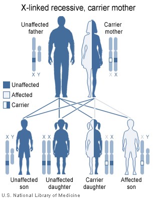
Those with protanopia, deuteranopia, protanomaly, and deuteranomaly have difficulty with discriminating red and green hues. Genetic red-green color blindness affects men much more often than women, because the genes for the red and green color receptors are located on the X chromosome, of which men have only one and women have two. Such a trait is called sex-linked. Females (46, XX) are red-green color blind only if both their X chromosomes are defective with a similar deficiency, whereas males (46, XY) are color blind if their single X chromosome is defective.
The gene for red-green color blindness is transmitted from a color blind male to all his daughters who are heterozygote carriers and are usually unaffected. In turn, a carrier woman has a fifty percent chance of passing on a mutated X chromosome region to each of her male offspring. The sons of an affected male will not inherit the trait from him, since they receive his Y chromosome and not his (defective) X chromosome. Should an affected male have children with a carrier or colorblind woman, their daughters may be colorblind by inheriting an affected X chromosome from each parent.
Because one X chromosome is inactivated at random in each cell during a woman's development, it is possible for her to have four different cone types, as when a carrier of protanomaly has a child with a deuteranomalic man. Denoting the normal vision alleles by P and D and the anomalous by p and d, the carrier is PD pD and the man is Pd. The daughter is either PD Pd or pD Pd. Suppose she is pD Pd. Each cell in her body expresses either her mother's chromosome pD or her father's Pd. Thus her red-green sensing will involve both the normal and the anomalous pigments for both colors. Such women are tetrachromats, since they require a mixture of four spectral lights to match an arbitrary light.
Blue-yellow color blindness
Those with tritanopia and tritanomaly have difficulty with discriminating blue and yellow hues.
Color blindness involving the inactivation of the short-wavelength sensitive cone system (whose absorption spectrum peaks in the bluish-violet) is called tritanopia or, loosely, blue-yellow color blindness. The tritanopes neutral point occurs near a yellowish 570 nm; green is perceived at shorter wavelengths and red at longer wavelengths. Mutation of the short-wavelength sensitive cones is called tritanomaly. Tritanopia is equally distributed among males and females. Jeremy H. Nathans (with the Howard Hughes Medical Institute) proved that the gene coding for the blue receptor lies on chromosome 7, which is shared equally by men and women. Therefore it is not sex-linked. This gene does not have any neighbor whose DNA sequence is similar. Blue color blindness is caused by a simple mutation in this gene. (2006, Howard Hughes Medical Institute).
Prevalence
Color blindness affects a significant number of people, although exact proportions vary among groups. In Australia, for example, it occurs in about 8 percent of males and only about 0.4 percent of females.[18] Isolated communities with a restricted gene pool sometimes produce high proportions of color blindness, including the less usual types. Examples include rural Finland, Hungary, and some of the Scottish islands. In the United States, about 7 percent of the male population – or about 10.5 million men – and 0.4 percent of the female population either cannot distinguish red from green, or see red and green differently (Howard Hughes Medical Institute, 2006). It has been found that more than 95 percent of all variations in human color vision involve the red and green receptors in male eyes. It is very rare for males or females to be "blind" to the blue end of the spectrum.
| Men | Women | Total | References | |
|---|---|---|---|---|
| Overall | — | — | — | |
| Overall (United States) | — | — | 1.30% | [19] |
| Red-green (Overall) | 7 to 10% | — | — | [20][19] |
| Red-green (Caucasians) | 8% | — | — | [21] |
| Red-green (Asians) | 5% | — | — | [21] |
| Red-green (Africans) | 4% | — | — | [21] |
| Monochromacy | — | — | — | |
| Rod monochromacy (disfunctional, abnormally shaped or no cones) | 0.00001% | 0.00001% | — | [22] |
| Dichromacy | 2.4% | 0.03% | — | [22] |
| Protanopia (L-cone absent) | 1% to 1.3% | 0.02% | — | [19][22] |
| Deuteranopia (M-cone absent) | 1% to 1.2% | 0.01% | — | [19][22] |
| Tritanopia (S-cone absent) | 0.001% | 0.03% | — | [22] |
| Anomalous Trichromacy | 6.3% | 0.37% | — | [22] |
| Protanomaly (L-cone defect) | 1.3% | 0.02% | — | [22] |
| Deuteranomaly (M-cone defect) | 5.0% | 0.35% | — | [22] |
| Tritanomaly (S-cone defect) | 0.01% | 0.01% | — | [22] |
Diagnosis

The Ishihara color test, which consists of a series of pictures of colored spots, is the test most often used to diagnose red-green color deficiencies. A figure (usually one or more Arabic digits) is embedded in the picture as a number of spots in a slightly different color, and can be seen with normal color vision, but not with a particular color defect. The full set of tests has a variety of figure/background color combinations, and enable diagnosis of which particular visual defect is present. The anomaloscope, described above, is also used in diagnosing anomalous trichromacy.
However, the Ishihara color test is criticized for containing only numerals and thus not being useful for young children, who have not yet learned to use numerals. It is often stated that it is important to identify these problems as soon as possible and explain them to the children to prevent possible problems and psychological traumas. For this reason, alternative color vision tests were developed using only symbols (square, circle, car).
Most clinical tests are designed to be fast, simple, and effective at identifying broad categories of color blindness. In academic studies of color blindness, on the other hand, there is more interest in developing flexible tests to collect thorough datasets, identify copunctal points, and measure just noticeable differences.[23]
Treatment and management
There is generally no treatment to cure color deficiencies. However, certain types of tinted filters and contact lenses may help an individual to distinguish different colors better. Optometrists can supply a singular red-tint contact lens to wear in the dominant eye. This may enable the wearer to pass color blindness tests for certain occupations. The effect of wearing such a device is akin to wearing red/blue 3D glasses and can take some getting used to as certain wavelengths can "jump" out and be overly represented. Additionally, computer software has been developed to assist those with visual color difficulties.
The Gnome Desktop provides colorblind accessibility using the gnome-mag and the libcolorblind software. Using a gnome applet, the user may switch a color filter on and off choosing from a set of possible color transformations that will displace the colors in order to disambiguate the colors. The software enables, for instance, a color blind person to see the numbers in the ishihara test.
The National Eye Institute is doing research into treating/curing color blindness, and it is now required to donate 5% of its resources to this cause under instruction of the National Institutes of Health.
Design implications of color blindness
Color codes present particular problems for color blind people as they are often difficult or impossible for color blind people to understand.
Good graphic design avoids using color coding or color contrasts alone to express information, as this not only helps color blind people, but also aids understanding by normally sighted people. The use of Cascading Style Sheets on the world wide web allows pages to be given an alternative color scheme for color-blind readers. This color scheme generator helps a graphic designer see color schemes as seen by eight types of color blindness. For an example of a map that could present a significant problem to a color blind reader, see this graphic from a recent New York Times article. The typical red-green color blind reader will find the green sections of the map nearly indistinguishable from the orange, rendering the graphic unreadable.
Designers should take into account that color-blindness is highly sensitive to differences in material. For example, a red-green colorblind person who is incapable of distinguishing colors on a map printed on paper may have no such difficulty when viewing the map on a computer screen or television. In addition, some color blind people find it easier to distinguish problem colors on artificial materials, such as plastic or in acrylic paints, than on natural materials, such as paper or wood. Thirdly, for some color blind people, color can only be distinguished if there is a sufficient "mass" of color: thin lines might appear black while a thicker line of the same color can be perceived as having color.
When the need to process visual information as rapidly as possible arises, for example in a train or aircraft crash, the visual system may operate only in shades of grey, with the extra information load in adding color being dropped. This is an important possibility to consider when designing, for example, emergency brake handles or emergency phones.
Due to this inability to recognize colors such as red and green, some countries (e.g., Singapore prior to the 1990s or Romania even to the present day) have refused to grant individuals with color blindness driving licenses. In Romania there is an ongoing effort to remove the legal restrictions that prohibit its colorblind citizens from getting drivers' licenses.[24]
Misconceptions and compensations
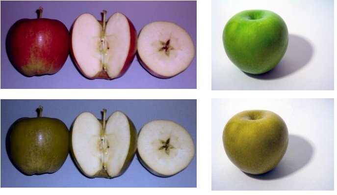
Color blindness is not the swapping of colors in the observer's eyes. Grass is never red, and stop signs are never green. The color impaired do not learn to call red "green" and vice versa. However, dichromats often confuse red and green items. For example, they may find it difficult to distinguish a Braeburn from a Granny Smith and in some cases, the red and yellow of a traffic light without other clues (e.g., shape or location). This is demonstrated in this simulation of the two types of apple as viewed by a trichromat or by a dichromat.
Anomalous Trichromats are often able to readily spot camouflage clothing, netting, and paint that has been designed for individuals with color-normal vision. They tend to learn to see texture and shape. This lets them see through some camouflage patterns.[1]
Traffic light colors are confusing to some dichromats: there is insufficient apparent difference between the red and amber and sodium street lamps and the green can be confused with a grubby white lamp. This is a risk factor on a high-speed undulating road where angular cues can't be used. British Rail color lamp signals use more easily identifiable colors: the red is really blood red, the amber is quite yellow and the green is a bluish color. Most British road traffic lights use a white rectangle; dichromats simply look for the position of the light within the rectangle (top, middle or bottom; British traffic lights are never mounted horizontally).
Color blindness almost never means complete monochromatism. In almost all cases, color blind people retain blue-yellow discrimination, and most color blind individuals are anomalous trichromats rather than complete dichromats. In practice this means that they often retain a limited discrimination along the red-green axis of color space although their ability to separate colors in this dimension is severely reduced.
It should also be noted that even though some people are unable to see some or maybe even any of the numbers in (e.g., red-green) color blindness tests, they might still be able to tell the difference between the colors in their everyday lives.
See also
- Cerebral achromatopsia
- Category:Articles with images not understandable by color blind users
References
- ↑ 1.0 1.1 Dalton J, 1798 "Extraordinary facts relating to the vision of colours: with observations" Memoirs of the Literary and Philosophical Society of Manchester 5 28-45
- ↑ Morgan MJ, Adam A, Mollon JD. "Dichromats detect colour-camouflaged objects that are not detected by trichromats." Proc Biol Sci. 1992 Jun 22;248(1323):291-5. PMID 1354367.
- ↑ Onishi, A., Koike, S., Ida, M., Imai, H., Shichida, Y., Takenaka, O., et al. (1999). Dichromatism in macaque monkeys. Nature, 402,139-140.
- ↑ Jordan G, Mollon JD.,A study of women heterozygous for colour deficiencies. Vision Res. 1993 Jul;33(11):1495-508. Jameson K.A., Richer color experience in observer with multiple photopigment opsin genes, Psychomomic Bulletin & Review, 2001, 8(2),244-261
- ↑ Sharpe, LT; Stockman A, Jägle H, Nathans J (1999). "Opsin genes, cone photopigments, color vision and color blindness". in Gegenfurtner KR, Sharpe LT. Color Vision: From Genes to Perception. Cambridge University Press. ISBN 9780521004398. http://books.google.com/books?id=4zQMQLLVkFYC.
- ↑ 6.0 6.1 "Color Blindness." University of Illinois Eye Center, Department of Ophthalmology and Visual Sciences. Accessed September 29, 2006.
- ↑ Kokotailo R, Kline D. "Congenital Colour Vision Deficiencies." University of Calgary, Department of Psychology, Vision & Aging Lab. Accessed September 29, 2006.
- ↑ 8.0 8.1 8.2 8.3 "Guidelines: Color Blindness." Tiresias.org. Accessed September 29, 2006.
- ↑ 9.0 9.1 9.2 9.3 9.4 9.5 9.6 9.7 9.8 9.9 Cassin, B. and Solomon, S. Dictionary of Eye Terminology. Gainsville, Florida: Triad Publishing Company, 1990.
- ↑ Spring, Kenneth R.; Matthew J. Parry-Hill; Thomas J. Fellers; Michael W. Davidson. "Human Vision and Color Perception". Florida State University. Retrieved on 2007-04-05.
- ↑ Paul S. Hoffman. "Accommodating Color Blindness" (PDF). Retrieved on 2007-04-05.
- ↑ Neitz, Maureen E., PhD. "Severity of Colorblindness Varies". Medical College of Wisconsin. Retrieved on 2007-04-05.
- ↑ 13.0 13.1 Byrne A, Hilbert D. "A Glossary of Color Science." Originally published in Readings on Color, Volume 2: The Science of Color. (MIT Press, 1997). Accessed November 7, 2006.
- ↑ *Weiss AH, et al 1989. "Blue cone monochromatism" J Pediatr Ophthalmol Strabismus. 1989; 11: 315-7
- ↑ 15.0 15.1 15.2 Kalloniatis, Michael and Luu, Charles. "Psychophysics of Vision: The Perception of Color". Retrieved on 2007-04-02.
- ↑ Montgomery, Ted M., O.D.. "The Macula". Retrieved on 2007-04-02.
- ↑ "Disease-causing Mutations and protein structure". UCL Biochemistry BSM Group. Retrieved on 2007-04-02.
- ↑ "Colour blindness". Better Health Channel. Retrieved on 2007-04-02.
- ↑ 19.0 19.1 19.2 19.3 "Prevalence and Incidence of Color blindness". WrongDiagnosis.com (2008-06-22). Retrieved on 2008-07-10.
- ↑ Color Blindness: More Prevalent Among Males
- ↑ 21.0 21.1 21.2 Masataka Okabe, Kei Ito (2008-02-15). "Colorblind Barrier Free". J*Fly. Retrieved on 2008-07-10.
- ↑ 22.0 22.1 22.2 22.3 22.4 22.5 22.6 22.7 22.8 http://www.colorfield.com/ref/types.html
- ↑ HubMed Abstracts
- ↑ "Petition to European Union on Colorblind’s condition in Romania". Retrieved on 2007-08-21.
^ a b c d e f g h i http://www.colorfield.com/ref/types.html[dead link]
have to be
^ a b c d e f g h i http://www.colorfilter.com/ref/types.html
Bibliography
- Smal, James; Hilbert, D.S. (1997). Readings on Color, Volume 2: The Science of Color (2nd ed. ed.). Cambridge, Massachusetts: MIT Press. ISBN 0-262-52231-4.
- Kaiser, Peter K.; Boynton, R.M. (1996). Human Color Vision (2nd ed. ed.). Washington, DC: Optical Society of America. ISBN 1-55752-461-0.
- Wyszecki, Günther; Stiles, W.S. (2000). Color Science: Concepts and Methods, Quantitative Data and Formulae (2nd edition ed.). places: Wiley-Interscience. ISBN 0-471-39918-3.
- McIntyre, Donald (2002). Colour Blindness: Causes and Effects. UK: Dalton Publishing. ISBN 0-9541886-0-8.
- Shevell, Steven K. (2003). The Science of Color (2nd ed. ed.). Oxford, UK: Optical Society of America. pp. 350. ISBN 0-444-512-519.
External links
- Color Blindness Examples
- Deuteranope Color Blindness Examples (Most common form of Red/Green)
- Color vision deficiency tests and facts
- Vischeck - Simulates colorblind vision.
- Colorblind Barrier-Free, contains a proposal for dichromacy safe palette.
- Online Color Blind Selftest
- WhatColor - Naming on-screen colors, handy tool for the color blind (Windows).
- Colorblind Helper - Naming colors in the world, turns iPhone into handheld color blind tool (iPhone).
- Colorblind Web Page Filter - View Web sites like a color blind person would see them.
- Color Universal Design Organization in Japan (CUDO) - How to create the Color Universal Design . Japanese Visual Science Groupe.
- Daltap The Daltonism Appliance: A handy tool for the color blind.
- ColorBlindExt Firefox add on for colorblind people
|
|||||||||||
|
||||||||||||||||||||||||||||||||||||||||||||||