Clavicle
- This article uses some professional terms to describe relative positions and directions. See Anatomical terms of location for more detailed information.
| Bone: Clavicle | |
|---|---|
 |
|
| Gray's | subject #49 200 |
| MeSH | Clavicle |
In human anatomy, the clavicle or collar bone is classified as a long bone that makes up part of the shoulder girdle (pectoral girdle). It receives its name from the Latin clavicula ("little key") because the bone rotates along its axis like a key when the shoulder is abducted. (This movement is palpable with the opposite hand). In some people, particularly females who may have less fat in this region, the location of the bone is clearly visible as it creates a bulge in the skin.
Contents |
Overview
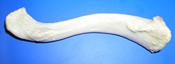  |
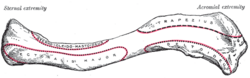
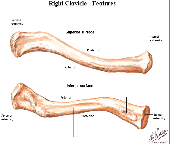
| Right clavicle - from below, and from above. |
 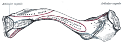 |
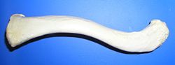
| Left clavicle - from above, and from below. |
The clavicle is a doubly curved short bone that connects the arm (upper limb) to the body (trunk), located directly above the first rib. It acts as a shunt to keep the scapula in position so the arm can hang freely. Medially, it articulates with the manubrium of the sternum (breast-bone) at the sternoclavicular joint. At its lateral end it articulates with the acromion of the scapula (shoulder blade) at the acromioclavicular joint. It has a rounded medial end and a flattened lateral end.
From the roughly pyramidal sternal end, each clavicle curves laterally and posteriorly for roughly half its length. It then forms a smooth posterior curve to articulate with a process of the scapula (acromion). The flat, acromial end of the clavicle is broader than the sternal end. The acromial end has a rough inferior surface that bears prominent lines and tubercles. These surface features are attachment sites for muscles and ligaments of the shoulder.
Functions
The clavicle serves several functions:
- It serves as a rigid support from which the scapula and free limb are suspended. This arrangement keeps the upper limb (arm) away from the thorax so that the arm has maximum range of movement.
- Covers the cervicoaxillary canal (passageway between the neck and arm), through which several important structures pass.
- Transmits physical impacts from the upper limb to the axial skeleton.
Even though it is classified as a long bone, the clavicle has no medullary (bone marrow) cavity like other long bones. It is made up of spongy (cancellous) bone with a shell of compact bone. It is a dermal bone derived from elements originally attached to the skull.
Attachments
Muscles and ligaments that attach to the clavicle include:
| Attachment on clavicle | Muscle/Ligament | Other attachment |
| Superior surface and anterior border | Deltoid muscle | deltoid tubercle, anteriorly on the lateral third |
| Superior surface | Trapezius muscle | posteriorly on the lateral third |
| Inferior surface | Subclavius muscle | subclavian groove |
| Inferior surface | Conoid ligament (the medial part of the coracoclavicular ligament) | conoid tubercle |
| Inferior surface | Trapezoid ligament (the lateral part of the coracoclavicular ligament) | trapezoid line |
| Anterior border | Pectoralis major muscle | medial third (rounded border) |
| Posterior border | Sternocleidomastoid muscle (clavicular head) | superiorly, on the medial third |
| Posterior border | Sternohyoid muscle | inferiorly, on the medial third |
| Posterior border | Trapezius muscle | lateral third |
Development
The clavicle is the first bone to begin the process of ossification (laying down of minerals onto a preformed matrix) during development of the embryo, during the 5th and 6th weeks of gestation. However, it is one of the last bones to finish ossification, at about 21-25 years of age. It forms by intramembranous ossification. It consists of a mass of cancellous bone surrounded by a compact bone shell. The cancellous bone forms via two ossification centres, one medial and one lateral, which fuse later on. The compact forms as the layer of fascia covering the bone stimulates the ossification of adjacent tissue. The resulting compact bone is known as a periosteal collar.
Common clavicle injuries
- acromioclavicular dislocation
- sternoclavicular dislocations
- clavicle fractures
- osteolysis
- degeneration of the clavicle
Additional images
See also
- Clavicle fracture
- Bone terminology
- Terms for anatomical location
- The collarbones are sometimes partly or completely absent in cleidocranial dysostosis
- Wishbone or furcula - the fused clavicles of most birds
References
- Chung, Kyung. Board Review Series: Gross Anatomy, 4th edition.
- Moore, Keith L. and Arthur F. Dalley. Clinically Oriented Anatomy, 4th edition.
- Stedman's Medical Dictionary, 27th edition.
- Mosby's Medical, Nursing, and Allied Health Dictionary, 5th ed.
- Title of a song played by Alkaline Trio
External links
|
|||||||||||||||||