Ascomycota
| Ascomycota | ||||||
|---|---|---|---|---|---|---|
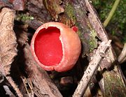 Sarcoscypha austriaca
|
||||||
| Scientific classification | ||||||
|
||||||
| Subphyla/Classes | ||||||
|
Ascomycota is a Division/Phylum of Fungi, and subkingdom Dikarya, whose members are commonly known as the Sac Fungi. They are the largest phylum of the kingdom Fungi, with over 30,000 species.[2] Characteristically, when reproducing sexually, they produce nonmotile spores in a distinctive type of microscopic cell called an "ascus" (from Greek: ἀσκός (askos), meaning "sac" or "wineskin"). These spores are called ascospores. However, some members of the Ascomycota do not reproduce sexually and do not form asci or ascospores. These members are assigned to Ascomycota based upon morphological and/or physiological similarities to ascus-bearing taxa, and in particular by phylogenetic comparisons of DNA sequences.[3][4]
This monophyletic grouping is an extremely significant and successful group of organisms. Familiar examples of sac fungi include morels, truffles, brewer's yeast and baker's yeast, Dead Man's Fingers, cup fungi, and the majority of lichens (loosely termed "ascolichens") such as Cladonia. Many plant-pathogenic fungi belong to the Ascomycota. Commonly seen examples include apple scab, ergot, black knot, and the powdery mildews. Species of ascomycetes are also popular in the laboratory. Sordaria fimicola, Neurospora crassa and several species of yeasts are used in many genetics and cell biology experiments. Penicillium species on cheeses and in the antibiotic industry are examples of asexual taxa, otherwise known as anamorphs, that belong in the Ascomycota. Prior to definitive phylogenetic research, molds such as Penicillium were sometimes classified in an artificial phylum, called the Deuteromycota.
Ascomycetes versus Ascomycota [4]
In the past, before the recognition of the fungal kingdom, the sac fungi were considered to be a Class, not a Phylum. The original collective term for them was "Ascomycetes", a label first coined in the 1800s for a rankless nonlichenized taxon based upon the presence of asci. "Ascomycetes" was soon used to include lichenized taxa, and became the standard term, at the class level, for all ascus-bearing species, just as the term "Basidiomycetes" became used for their basidium-bearing counterparts. Elevation of the taxonomic rank of the Ascomycetes resulted in the names Ascomycetae, Ascomycotina, and finally Ascomycota. The names Ascomycota, Ascomycetes, etc. are based upon the term "ascus". Together, the Ascomycota and the Basidiomycota form the subkingdom Dikarya. The more familiar term, Ascomycetes, is still loosely used, e.g. at fungal forays it is often said of a fungus, such as Peziza, "It is an ascomycete, not a basidiomycete" in reference to their sexual reproductive mode. The terms are further abbreviated to "ascos" and "basidos" which are not officially sanctioned technical names.
Modern classification of Ascomycota
There are 3 subphyla that are described and named:
- The subphylum Pezizomycotina is the largest subphylum and contains all the Ascomycota that produce ascocarps (fruiting bodies), except for one genus, Neolecta, in the Taphrinomycotina. Therefore, it includes virtually all macroscopic "ascos" such as truffles, ergot, ascolichens, cup fungi (discomycetes), pyrenomycetes, lorchels, and caterpillar fungus[3], as well as many microscopic fungi, e.g. powdery mildews, ring worm fungi, chalkbrood fungus[4], Laboulbeniales, and most black molds around sinks and tubs. The older named taxon Euascomycetes is roughly equivalent.
- The subphylum Saccharomycotina comprises most of the "true" yeasts, such as baker's yeast and Candida which are in general single-celled, or short chains of cells, and reproduce vegetatively by budding rather than by the production of hyphae. As a result, most were classified in a vaguely defined taxon with the older name Hemiascomycetes.
- The subphylum Taphrinomycotina includes a disparate group of Ascomycota and were only recognized as a distinctive group after the advent of molecular (DNA) analyses. The group is basal to the other subphyla and hence is considered to be more primitive. Consequently the taxon was originally named Archiascomycetes alternatively spelled Archaeascomycetes. It includes both hyphal fungi (Neolecta, Taphrina), and fission yeasts Schizosaccharomyces and the peculiar mammalian lung parasite, Pneumocystis that was originally believed to be a protozoan.
Evidence from ribosomal RNA gene sequencing of soils indicates that there likely a fourth previously unknown subphylum of Ascomycota (loosely termed Soil Clone Group I -SCGI), that has never been described via cultures or fruitbodies. SCGI organisms are only known from DNA sequences but have been shown to occur in soils worldwide by Schadt et al. (2003) [5] and Porter et al. (2007) [6]. Placement of this group based on rRNA gene sequencing indicates that they may fall between the Taphriomycotina and the Saccharomycotina, however phenotypic characteristics and environmental roles and significance remain unknown.
Commonly used but obsolete morphologically defined class names
Several obsolete class names, based upon morphology, are still used in informal or introductory discussions. Among those based upon the sexual fruitbodies (teleomorphs) are: the Discomycetes which included all species forming apothecia; the Pyrenomycetes which included all sac fungi that formed perithecia or even pseudothecia, or any structure approaching these morphological structures; and the Plectomycetes which included those Ascomycota that formed cleistothecia. Hemiascomycetes included the yeasts and yeast-like fungi that are now split between Saccharomycotina and Taphrinomycotina, while the Euascomycetes covered the rest of the Ascomycota, now in the Pezizomycotina and Neolecta in the Taphrinomycotina.
Some Ascomycota never reproduce sexually, or are not known to produce asci. These are sometimes called "Mitosporic Ascomycota" because of the production of conidia otherwise known as mitospores, and other asexual structures, all collectively called anamorphic taxa. In some classifications these would have been placed in a separate artificial phylum, the Deuteromycota (also known as Fungi Imperfecti). Molecular analyses can now be used to place these genera and species among ascus-bearing taxa (if they are Ascomycota), or amongst other phyla such as the Basidiomycota (if related to them). No mitosporic taxa have been found that form a phylum distinct from the other major phyla of Fungi. Anamorphs are discussed below. However, it is extremely difficult in the absence of molecular analysis to link most anamorphic (asexual morphs) fungi to their teleomorphs (sexual counterparts). There are over 250 years of names available (since Linnaeus' Species Plantarum, 1753) for both asexual and sexual components of the same fungi. For instance the sexual form of the kerosene fungus is known as Amorphotheca resinae while the asexual stage is called Hormoconis resinae. Most anamorphic fungi are Ascomycota, and therefore the obsolete classification of the Deuteromycota is largely that of Ascomycota anamorphs.
The Deuteromycota were classified as Coelomycetes if they produced their conidia in small seed-like, fly-speck sized, flask-shaped conidiomata, or structures resembling or approximating them in structure. The Hyphomycetes were those species where the conidiophores (i.e. the hyphae which carry conidia-forming cells on the end) are free or loosely organized. They are mostly isolated but sometimes also appear as bundles of cells aligned in parallel (described as synnematal) or as cushion-shaped masses (described as sporodochial).
Physical make-up
The adjective which describes these fungi is "ascomycetous". The majority of ascomycetous fungi grow as a thallus, called a mycelium, consisting of many hyphae which are microscopic multi-branched filaments. If the hyphae of some typical mycelia were laid end to end, they could reach a length of several kilometers. Ascomycota typically produce great numbers of asci at any one time, and these may be contained in a multicellular, often readily visible structure called an "ascocarp" (also called an "ascoma", the fruiting body of ascomycetes). Many exceptions to the structure described above occur, for example in one extreme these fungi are single celled yeasts, and there is no mycelium, no fruitbody, and the entire cell is converted into an ascus in such ascomycetous yeasts such as baker's Yeast (Saccharomyces cerevisiae).
In the case of lichenized species, the thallus of the fungus defines the shape of the symbiotic colony. Other Ascomycota are dimorphic, which can mean that they can appear either in single- or multi-cellular form. Other species are pleomorphic, exhibiting multiple asexual forms (i.e. anamorphs detailed below) as well as a sexual form (a teleomorph). The ascoma come in multiple forms: cup-shaped, club-shaped, potato-like, spongy, seed-like, oozing and pimple-like, coral-like, nit-like, golf-ball-shaped, perforated tennis ball-like, cushion-shaped, plated and feathered in miniature (Laboulbeniales), microscopic classic Greek shield-shaped, stalked or sessile, solitary or clustered, etc. They can be fleshy or carbonaceous (like charcoal), leathery, rubbery, gelatinous, slimy, powdery, or cob-web-like, etc. They come in multiple colors such as red, orange, yellow, and rarely green or blue, although brown or black are more common.
Except for lichens, the mycelium (if produced) is usually inconspicuous because it is subterranean or embedded in the substrate, and only the ascoma is seen in season. But spectacular, bizarre, or otherwise noteworthy exceptions occur. Many ascomatous fungi have melanized hyphal walls (referred to as dematiaceous walls) and therefore are black or brown. Black spots on bathroom caulking are often colonies of Ascomycota, e.g. Cladosporium. Many molds that grow on spoiled foods are Ascomycota, and therefore the pellicles or skins that develop on jams, juices, and other foods in containers at home are in fact the thalli of Ascomycota (occasionally Mucoromycotina, and almost never Basidiomycota). Sooty molds that develop on plants, especially in the tropics are the thalli of many species.
Sometimes it is the mass of asci or ascus-like cells, or conidia or yeast cells that are the conspicuous elements. Pneumocystis species fill lung cavities causing a form of pneumonia (visible in x-rays). Ascosphaera cysts (asci) fill honey bee larvae and pupae making them appear mummified and chalk-like, hence the name "chalkbrood". Free living yeasts form yeast colonies. Excessive Candida yeast growth in the mouth or vagina is called "thrush" or candidiasis.
The cell walls of these fungi are almost always formed of Chitin and β-Glucans; individual cells are formed from divisions of the hyphae called "septa". These give stability to the hyphae and prevent a great loss of cytoplasm in the event that the cell membrane should be locally damaged. Mostly the cell divisions are centrally perforated, so they have a small opening in the middle, through which cytoplasm and also nuclei can move more or less freely throughout the system of hyphae. Often hyphae have only one nucleus per cell, and are therefore described as uninucleate, but some ascomycetous fungi can also be multinucleate at times.

Metabolism
Like most fungi the Ascomycota principally digest living or dead biomass. To achieve this, they secrete into their surroundings powerful digestive enzymes which break down organic substances into small molecules, which are then absorbed through the cell wall. Many species live on dead plant material such as fallen leaves, twigs, or logs. Others attack plants, animals, or other fungi as parasites and derive their metabolic energy, as well as all the nutrients they need, from the cell tissue of their hosts. Especially in this group extreme specialization appears; for instance certain species of Laboulbeniales attack only one particular leg of one particular insect species. The Ascomycota also often take up symbiotic relationships – for instance some combine with green algae or cyanobacteria, from which they obtain photosynthetic nutrients, to form lichens; others form symbioses with tree roots as mycorrhizal fungi. There are also carnivorous fungi, which have developed hyphal traps in which they can catch small protists such as amoebae, as well as roundworms (Nematoda), rotifers, tardigrades, and small arthropods such as springtails (Collembola).
Through their long evolutionary history the Ascomycota have developed the capability to break down almost every organic substance. Unlike most organisms they are able to use their own enzymes to digest plant cellulose and the lignin contained in wood. Collagen, an abundant structural protein in animals, and keratin (which hair is made of), can also serve as food sources. Exotic examples are given by the ascomycete Aureobasidium pullulans, which metabolizes wall paint, and the kerosene fungus Amorphotheca resinae, which (to the misfortune of the airline industry) feeds on aircraft fuel, and in tropical regions sometimes blocks fuel pipes. Others resist osmotic stress to grow on salted fish, and a few live in water.

Distribution and living environment
The Ascomycota are present in all land ecosystems worldwide – they even occur in Antarctica – and their spores and hyphal fragments are distributed through the atmosphere and fresh water environments, as well as ocean beaches and tidal zones. The distribution of individual species is very variable: some are found on all continents, while for example the white truffle Tuber magnatum, which is much sought after for culinary purposes, only appears in isolated locations in Italy and France. Plant parasitic species are often restricted by their host distributions. Cyttaria is only found on Nothofagus (Southern Beech) in the Southern Hemisphere.
Reproduction
Asexual reproduction
Asexual reproduction is the dominant form of propagation in the Ascomycota, and is responsible for the rapid expansion of these fungi into areas which were previously not colonized. It occurs through reproductive structures, the "conidia," which are genetically identical to the parent and mostly have just one nucleus. They are also called "mitospores" due to the way they are generated through the cellular process of mitosis. They are generally formed on the ends of specialized hyphae, the "conidiophores". Depending on the species they may be dispersed by wind or water, or also by animals.
Asexual spores
In order to further classify the Ascomycota in the asexual stages, it is important to consider the spores, which can be distinguished by colour, form and the way they are separated into cells. The most frequent types are the single-celled spores which are designated amerospores. If the spore is divided into two by a cross-wall (septum), it is a didymospore.
Conidiospores of Trichoderma aggressivum, Diameter approx. 3µm
|

Conidiophores of molds of the genus Aspergillus, conidiogenesis is blastic-phialidic
|

Conidiophores of Trichoderma harzianum, conidiogenesis is blastic-phialidic
|

Conidiophores of Trichoderma fertile with vase-shaped phialides and newly formed conidia on their ends (bright points)
|
When there are two or more cross-walls the classification depends on the shape. If the septa are transversal, like the rungs of a ladder, it is a phragmospore whilst if they form a net-like structure it is a dictyospore. In staurospores ray-like "arms" radiate from a central body; in others (helicospores) the entire spore is wound up in a spiral like a spring. Finally very long worm-like spores, of which the ratio length:diameter is more than 15:1, are called scolecospores.
Conidiogenesis and dehiscence
One distinguishes:
- acervular conidiomata, or acervuli, which develop in the host and can thus be:
- subcuticular, lying under the outer layer of the plant (the cuticle),
- intraepidermal, inside the outer cell layer (the epidermis),
- subepidermal, under the epidermis, or
- deeper inside the host.
- Mostly they develop a flat layer of relatively short conidiophores which then produce masses of spores. The increasing pressure finally leads to the splitting of the epidermis and cuticle and so allows the conidia to escape.
- pycnidial conidiomata or pycnidia, which unlike the acervuli form in the fungal tissue itself, and which are generally shaped like a bulging vase. The spores are released through a small opening at the apex, the ostiole.
Two further important characteristics of the anamorphs of the Ascomycota are the conidiogenesis, the fashion in which the spores are formed, and their dehiscence, i.e. how they separate from the parent structures. The former corresponds to Embryology in animals and plants and can be divided into two fundamental forms of development: blastic conidiogenesis, where the spore is already evident before it separates from the conidiogenic hypha which is giving rise to it, and thallic conidiogenesis, where first a cross-wall appears and then the thus created cell develops into a spore.
These two basic types can be further classified as follows.
- blastic-acropetal (repeated budding at the tip of the conidiogenic hypha, so that a chain of spores is formed with the youngest at the tip),
- blastic-synchronous (simultaneous spore formation from a central cell, sometimes with secondary acropetal chains forming from the initial spores),
- blastic-sympodial (repeated sideways spore formation from behind the leading spore, so that the oldest spore is at the main tip),
- blastic-annellidic (each spore separates and leaves a ring-shaped scar which is concentrically inside the scar left by the previous spore),
- blastic-phialidic (the spores arise and are ejected from the open ends of special conidiogenic cells called phialides which remain constant in length; an example is the anamorph of Penicillium),
- basauxic (where a chain of conidia, in successively younger stages of development, is emitted from the mother cell),
- blastic-retrogressive (spores separate off by formation of crosswalls near the tip of the conidiogenic hypha, which thus becomes progressively shorter),
- thallic-arthric (double cell walls split the conidiogenic hypha into cells which develop into short, cylindrical spores called arthroconidia; sometimes every second cell dies off, leaving the arthroconidia free),
- thallic-solitary (a large bulging cell separates from the conidiogenic hypha, forms internal walls, and develops to a phragmospore).
Essentially dehiscence can happen in two different ways. In the schizolytic variant a double dividing wall with a central lamella (layer) forms between the cells; the central layer dissolves to release the spores. In the case of rhexolytic dehiscence on the other hand the cell wall which joins the spores on the outside simply degenerates and sets free the conidia.
Heterocaryosis and parasexuality
A significant number of Ascomycota species either have no sexual stage or none is known. In spite of this, there are two ways in which they can conserve their genetic diversity: Heterocaryosis and Parasexuality.
The former happens simply through the merging of two hyphae belonging to different individuals, a process known as anastomosis. As a result there are more cell nuclei than normal in the mycelium and they come from genetically different parent organisms.
Parasexuality, on the other hand, refers to a phenomenon where two cell nuclei merge without any sexual process and the chromosome count is doubled. This involves a complex form of the type of cell division called mitosis, where there is crossing over or recombination, i.e. an exchange of genetic material between corresponding pairs of chromosomes. In sexual reproduction, in contrast, crossing over occurs only during meiosis. Finally the chromosome count will be restored to normal by haploidization, whereby the nucleus splits into two parts each having a single set of chromosomes, with each daughter genetically different from the original parents.
Sexual reproduction

Sexual reproduction in the Ascomycota is marked by a characteristic structure, the ascus, which distinguishes these fungi from all others. An ascus is a tube-shaped vessel, a meiosporangium, which contains the sexual spores produced by meiosis. The latter are called ascospores in contrast to the asexual conidiospores.
Apart from exceptions such as baker's Yeast (Saccharomyces cerevisiae), almost all fungi of the Ascomycota are haploid, so their nuclei only contain one set of chromosomes, which makes them especially susceptible to mutations. During sexual reproduction there is a diploid phase (with two sets of chromosomes), which as a rule is very short. Then meiosis occurs, generally very soon, so that the haploid state is re-established.
The formation of sexual spores
The sexual part of the life cycle commences when two suitable hyphae meet each other. These come from the same web of hyphae which can also generate asexual spores. The first deciding factor as to whether conjugation - that is, sexual merging - will occur, is whether the hyphae belong to the same organism, or whether they come from different individual fungi. Whilst many species are thoroughly capable of self-propagation, i.e. they are homothallic, others need non-identical partners and so are heterothallic. Besides this, the two hyphae in question must also belong to the same mating type. Mating types are a peculiarity of the fungi and correspond roughly to the sexes in plants and animals; however one species may have more than two mating types.
In the case of compatibility, gametangia form on the hyphae; these are the generative cells for the gametes, in which numerous nuclei gather. A very fine hypha, called the trichogyne, which grows out of one gametangium, now termed the ascogonium, makes a passage to a gametangium of the other individual, which is then the antheridium. Nuclei then pass from the antheridium (playing a 'male' role) to the ascogonium (playing a 'female' role).
Unlike the process in animals and plants, after the union of the cytoplasms of the two gametangia (plasmogamy), the merging of the nuclei (karyogamy) does not usually occur immediately. Instead, the nuclei which have migrated in from the antheridium pair up with the nuclei of the ascogonium, but remain separate next to their partners. With this the dikaryophase of the life cycle begins; during this time the pairs of nuclei repeatedly synchronously divide, so that a great number are produced. In all probability the dikaryophase is an evolutionary adaptation which serves to exploit the potential of sexual reproduction to the full in circumstances where it is a rare event for different individuals to meet each other. After the genetic raw material has been increased by repeated division, recombination will take place independently in each pair during meiosis, so that the greatest possible quantity of genetically different spores will arise. In the red algae (Rhodophyta) a similar solution to the corresponding problem evolved independently.
Next millions of new dinucleate hyphae, into each of which two nuclei migrate, emerge from the fertilized ascogonium. They are also called ascogenous or fertile. They are fed by ordinary uni- or mononucleate hyphae (with only one nucleus), which are also called sterile. The tissue of sterile and fertile hyphae now grows in many cases into a macroscopically visible fruiting body, the ascocarp, which may contain millions of fertile hyphae.

In the actual fruiting layer, the hymenium, the asci now appear. At one end of an ascogenous hypha, there develops a U-shaped hook, which points back opposite to the general growth direction. The two nuclei contained in the terminal cell then divide in such a way that the threads of their mitotic spindles run parallel, and thus two pairs of genetically different daughter nuclei arise, with one daughter of each pair near the point of the hook, and the other in the base part of the hypha. Then two parallel cross-walls appear, dividing the hypha into three sections: that at the point of the hook with one nucleus, that at the base of the original hypha with one nucleus, and the middle U-shaped part with two nuclei.
If the positioning in the fruiting layer is right, the karyogamic fusion of the nuclei finally takes place in the U-shaped cell, creating the diploid zygote. It lengthens to form an elongated tube-shaped or cylinder-shaped capsule, the actual ascus. Then meiosis occurs, giving rise to four haploid nuclei. This is almost always followed by a further mitotic division, so that the ascus ultimately has eight daughter nuclei. These become enclosed, together with some of the cell plasma, each by their own membranes, and generally with a hard cell wall. Thus the dissemination cells (the ascospores) develop, lying initially like peas in a pod inside the ascus. Later, when an appropriate opportunity presents itself, they are liberated.
Not having flagella, ascospores are disseminated in various other ways: some are spread by wind and with others the ripe ascus breaks open on contact with water to set free the spores. Certain species have evolved regular 'spore cannons' which can eject them up to 30 cm. away. When the spores reach a suitable substrate, they germinate, form new hyphae, and so restart their life cycle, which has come full circle.
The form of the ascus is important for classification and is divided into four basic types: unitunicate-operculate, unitunicate-inoperculate, bitunicate, or prototunicate. See the article on asci for further details.
Ecology
The Ascomycota fulfil a central role in most land-based ecosystems. They are important decomposers which break down such organic materials as dead leaves, twigs, fallen trees, etc. and help the detritivores (animals which live off this decomposing material) to obtain their nutrients. By processing substances like cellulose or lignin, which are otherwise difficult to exploit, they take on an important place in the natural nitrogen cycle and the carbon cycle.
Inversely the fruiting bodies of the Ascomycota provide food for a very diverse set of animals from insects and slugs and snails (Gastropoda) to rodents and larger mammals such as deer and wild boars.
Fungi of the Ascomycota are also known for their numerous symbiotic relationships with other organisms.
Lichens
Cross-section through the lichen Pseudevernia furfuracea with plainly visible layer of green algae under the surface
|

Pseudevernia furfuracea
|
Probably since early in their evolutionary history the Ascomycota have "domesticated" green algae (Chlorophyta), as well as occasionally other types of algae and cyanobacteria. Together they form the mutualistic associations known as lichens, which can survive in the least hospitable regions of the earth, including the Arctic, the Antarctic, deserts and mountaintops, and can withstand temperature extremes from -40°C to +80°C. While the photoautotrophic algal partner creates metabolic energy through photosynthesis, the fungus offers a stable supportive framework and protects from radiation and drying out. Around 42% of the Ascomycota (numerically about 18,000 species) form lichens, and almost all the fungal partners of lichens belong to the Ascomycota - the proportion of Basidiomycota is probably only two to three percent.
Mycorrhizal fungi and endophytes
Members of the Ascomycota make two particularly important types of relationship with plants: as mycorrhizal fungi and as endophytes. The former make symbiotic associations with the root systems of the plants, which for some trees, especially conifers, can be of vital importance, enabling the uptake of mineral salts from the soil. The fungal partner is in a much better position to absorb minerals due to its finely divided mycelium, whilst the plant provides it with metabolic energy in the form of photosynthetic products. Cases are even known where mycorrhizal fungi can transport nutrients from one plant to another, stabilizing the recipient. It is likely that mycorrhizal associations enabled the conquest of the land by plants - in any case the earliest known fossils of land plants have mycorrhizae.
Endophytes on the other hand live inside plants, especially in the stem and leaves, but generally do not damage their hosts. The exact nature of the relationship between endophytic fungus and host is not yet well understood, but it seems that this form of colonization can bestow a higher resistance against insects, roundworms (nematodes), and bacteria; also it can enable or augment the production of poisonous alkaloids, chemicals which can affect the health of plant-eating mammals.
Symbiotic relationships with animals
A series of Ascomycota species from the genus Xylaria are found in the nests of leafcutter ants and other fungus-growing ants of the tribe Attini and in the fungal gardens of termites (Isoptera). Since they do not generate fruiting bodies until the insects have left the nests, it is suspected that, as confirmed in several cases of Basidiomycota species, they may be cultivated.
On the other hand bark beetles (Scolytidae) are certainly important symbiotic partners. The female beetles transport the spores to new hosts in characteristic tucks in their skin, the mycetangia. There they eat tunnels in the wood, which lead into large chambers in which they lay their eggs. At this time the spores are released and give rise to hyphae which unlike the beetles can digest the wood. The beetle larvae feed on the fungus and after they have metamorphosed into the adult state they again carry spores with them to renew the cycle of infection. A well-known example of this is Dutch elm disease, caused by fungus Ophiostoma ulmi, being carried by the European elm bark beetle Scolytus multistriatus.
Importance for humans

Ascomycetes make many contributions to the good of humanity, and also have many ill effects.
Harmful interactions
One of their most harmful roles is as the agent of many plant diseases. For instance:
- Dutch Elm Disease, caused by the closely related species Ophiostoma ulmi and Ophiostoma novo-ulmi, has led to the death of many elms in Europe and North America.

- The originally Asian Cryphonectria parasitica is responsible for attacking Sweet Chestnuts (Castanea sativa), and virtually eliminated the once-widespread American Chestnut (Castanea dentata),
- A disease of Maize (Zea mays), which is especially prevalent in North America, is brought about by Cochliobolus heterostrophus.
- Taphrina deformans causes leaf curl of peach.
- Uncinula necator is responsible for the disease Powdery mildew, which attacks grapevines.
- Species of Monilia cause brown rot of stone fruit such as peaches (Prunus persica) and sour cherries (Prunus ceranus).
- Members of the Ascomycota such as Stachybotrys chartarum are responsible for fading of woollen textiles, which is a common problem especially in the tropics.
- Blue-green, red and brown moulds attack and spoil foodstuffs - for instance Penicillium italicum rots oranges.
- Cereals infected with Fusarium graminearum contain mycotoxins like deoxynivalenol (DON), which can lead to skin and mucous membrane lesions when eaten by pigs.
- Ergot (Claviceps purpurea) is a direct menace to humans when it attacks wheat or rye and produces highly poisonous and carcinogenic alkaloids, causing ergotism if consumed. Symptoms include hallucinations, stomach cramp, and a burning sensation in the limbs ("Saint Anthony's Fire").
- Aspergillus flavus, which grows on peanuts and other hosts, generates aflatoxin, which damages the liver and is highly carcinogenic.
- Candida albicans, a yeast which attacks the mucous membranes, can cause an infection of the mouth or vagina called thrush or candidiasis, and is also blamed for "yeast allergies".
- Fungi like Epidermophyton cause skin infections but are not very dangerous for people with healthy immune systems. However if the immune system is damaged they can be life-threatening; for instance, Pneumocystis jiroveci is responsible for severe lung infections which occur in AIDS patients.
Positive effects
On the other hand, ascus fungi have brought some important benefits to humanity.
- The most famous case may be that of the mould Penicillium chrysogenum (formerly Penicillium notatum), which, probably to attack competing bacteria, produces an antibiotic which, under the name of Penicillin, triggered a revolution in the treatment of bacterial infectious diseases in the 20th century.
- The medical importance of Tolypocladium niveum as an immunosuppressor can hardly be exaggerated. It excretes Ciclosporin, which, as well as being given during organ transplants to prevent rejection, is also prescribed for auto-immune diseases such as multiple sclerosis, although there is some doubt over the long-term side-effects of the treatment.

- Some ascomycete fungi can be altered relatively easily through genetic engineering procedures. They can then produce useful proteins such as insulin, human growth hormone, or TPa, which is employed to dissolve blood clots.
- The red bread mold Neurospora crassa is an important model organism in biology, of which the genome has now been fully sequenced.
- Baker's Yeast (Saccharomyces cerevisiae) is used to make bread, beer and wine, during which process sugars such as glucose or sucrose are fermented to make alcohol and carbon dioxide. In the case of bread-making, the alcohol evaporates and the carbon dioxide serves to make the dough rise.
- Enzymes of Penicillium camemberti play a role in the manufacture of the cheeses Camembert and Brie, while those of Penicillium roqueforti do the same for Gorgonzola, Roquefort and Stilton.
- In Asia Aspergillus oryzae is added to a pulp of soaked soya beans to make soy sauce.
- Finally, some members of the Ascomycota are eaten with relish; morels (Morchella) and truffles (Tuber) are some of the most sought-after fungus delicacies.
Notes
- a (Taylor, Spatafora & Berbee 1996) and reference 4.
- b The taxonomic system used here (based on reference 1) is only one among several; another authoritative one is given by references 2 & 3.
- d See reference 5.
Linked references
- All but the first 2 sections are translated from the German article.
- ↑ Cavalier-Smith, T. (1998). "A revised six-kingdom system of Life". Biol. Rev. Camb. Philos. Soc. 73 (3): 203–266. doi:. http://journals.cambridge.org/action/displayIssue?jid=BRE&volumeId=73&issueId=03#.
- ↑ Bisby, Guy Richard; Ainsworth, G. C.; Kirk, P. M.; Aptroot, André (2001). Ainsworth & Bisby's Dictionary of the fungi / by P. M. Kirk... [et al.]; with the assistance of A. Aptroot... [et al.]. Oxon: CAB International. ISBN 0-85199-377-X.
- ↑ Lutzoni F, et al (2004). "Assembling the fungal tree of life: progress, classification, and evolution of subcellular traits". Amer J Bot 91: 1446–1480. doi:10.3732/ajb.91.10.1446 (inactive 2008-06-25).
- ↑ James TY et al (2006). "Reconstructing the early evolution of Fungi using a six-gene phylogeny". Nature 443: 818–822. doi:10.1038/nature05110 (inactive 2008-06-25). PMID 17051209.
- ↑ Schadt et al., Science, 2003 [1]
- ↑ Porter et al., Molecular Phylogenetics and Evolution, 2007 [2]
Unlinked references
- Taylor, John W.; Joey Spatafora & Mary Berbee (March 11, 1996), "Ascomycota", Tree of Life, <http://tolweb.org/Ascomycota>.
- Ainsworth and Bisby's Dictionary of the Fungi, 9th Edition, see here for more details.
- See the Index Fungorum (Hierarchy Search) for a web search based on the previous reference.
- Outline of Ascomycota - 2001
- Palæos Fungi
- C. J. Alexopoulos, M. Blackwell, C. W. Mims: Introductory Mycology, 4th Ed., 1996, ISBN 0-471-52229-5
- B. Kendrick: "The Fifth Kingdom, 3rd Ed., 2001, Kapitel 4, ISBN 1-58510-022-6
- G. J. Krieglsteiner: Verbreitungsatlas der Großpilze Deutschlands (West), Volume 2: Schlauchpilze, Ulmer Verlag, 1993
- F. Breitenbach, J. Kränzlin: Pilze der Schweiz, Volume 1, Ascomycetes, Mykologia Luzern, 1984
- Anamorph-Teleomorph-Datenbank
- Fotografien einiger Fruchtkörper (Ascomata)
|
|||||||||||||