Artery
Arteries are blood vessels that carry blood away from the heart. All arteries, with the exception of the pulmonary and umbilical arteries, carry oxygenated blood.
The circulatory system is extremely important for sustaining life. Its proper functioning is responsible for the delivery of oxygen and nutrients to all cells, as well as the removal of carbon dioxide and waste products, maintenance of optimum pH, and the mobility of the elements, proteins and cells of the immune system. In developed countries, the two leading causes of death, myocardial infarction and stroke each may directly result from an arterial system that has been slowly and progressively compromised by years of deterioration. (See atherosclerosis).
Contents |
Description
The arterial system is the higher-pressure portion of the circulatory system. Arterial pressure varies between the peak pressure during heart contraction, called the systolic pressure, and the minimum, or diastolic pressure between contractions, when the heart expands and refills. This pressure variation within the artery produces the pulse which is observable in any artery, and reflects heart activity.[1]
Anatomy
The anatomy of arteries can be separated into gross anatomy, at the macroscopic level, and microscopic anatomy, which must be studied with the aid of a microscope.
Gross anatomy
- See also: Arterial tree
The arterial system of the human body is divided into systemic arteries, carrying blood from the heart to the whole body, and pulmonary arteries, carrying blood from the heart to the lungs.
Systemic arteries
- See also: Systemic circulation
- See also: Arterial tree
Systemic arteries are the arteries of the systemic circulation, which is the portion of the cardiovascular system which carries oxygenated blood away from the heart, to the body, and returns deoxygenated blood back to the heart.
Pulmonary arteries
- See also: Pulmonary circulation
Pulmonary arteries are the arteries of the pulmonary circulation, which is the portion of the cardiovascular system which carries deoxygenated blood away from the heart, to the lungs, and returns oxygenated blood back to the heart.

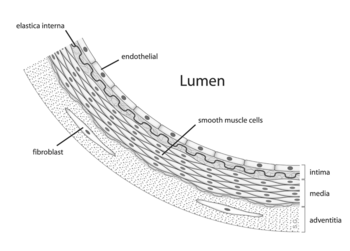
The outermost layer is known as the tunica externa formerly known as "tunica adventitia" and is composed of connective tissue. Inside this layer is the tunica media, or media, which is made up of smooth muscle cells and elastic tissue. The innermost layer, which is in direct contact with the flow of blood is the tunica intima, commonly called the intima. This layer is made up of mainly endothelial cells. The hollow internal cavity in which the blood flows is called the lumen.
Types of arteries
Pulmonary arteries
The pulmonary arteries carry deoxygenated blood that has just returned from the body to the heart towards the lungs, where carbon dioxide is exchanged for oxygen.
Systemic arteries
Systemic arteries can be subdivided into two types; muscular and elastic; according to the relative compositions of elastic and muscle tissue in their tunica media as well as their size and the makeup of the internal and external elastic lamina. The larger arteries >1cm diameter are generally elastic and the smaller ones 0.1-10mm tend to be muscular. Systemic arteries deliver blood to the arterioles, and then to the capillaries, where nutrients and gasses are exchanged.
The Aorta
The aorta is the root systemic artery. It receives blood directly from the left ventricle of the heart via the aortic valve. As the aorta branches, and these arteries branch in turn, they become successively smaller in diameter, down to the arteriole. The arterioles supply capillaries which in turn empty into venules. The very first branches off of the aorta are the coronary arteries, which supply blood to the heart muscle itself. These are followed by the branches off the aortic arch, namely the brachiocephalic artery, the left common carotid and the left subclavian arteries.
Arterioles
Arterioles, the smallest of the true arteries, help regulate blood pressure by the variable contraction of the smooth muscle of their walls, and deliver blood to the capillaries.
Arterioles and blood pressure
Arterioles have the greatest collective influence on both local blood flow and on overall blood pressure. They are the primary "adjustable nozzles" in the blood system, across which the greatest pressure drop occurs. The combination of heart output (cardiac output) and systemic vascular resistance, which refers to the collective resistance of all of the body's arterioles, are the principal determinants of arterial blood pressure at any given moment.
Capillaries
The capillaries are where all of the important exchanges happen in the circulatory system. The capillaries are a single cell in diameter to aid fast and easy diffusion of gases, sugars and other nutrients to surrounding tissues.
Functions of capillaries
To withstand and adapt to the pressures within, arteries are surrounded by varying thicknesses of smooth muscle which have extensive elastic and inelastic connective tissues.
The pulse pressure, i.e. Systolic vs. Diastolic difference, is determined primarily by the amount of blood ejected by each heart beat, stroke volume, versus the volume and elasticity of the major arteries.
Over time, elevated arterial blood sugar (see Diabetes Mellitus), lipoprotein cholesterol, and pressure, smoking, and other factors are all involved in damaging both the endothelium and walls of the arteries, resulting in atherosclerosis or Diabetes Mellitus.
History
Among the ancient Greeks, the arteries were considered to be "air holders" that were responsible for the transport of air to the tissues and were connected to the trachea. This was as a result of the arteries of the dead being found to be empty.
In medieval times, it was recognized that arteries carried a fluid, called "spiritual blood" or "vital spirits", considered to be different from the contents of the veins. This theory went back to Galen. In the late medieval period, the trachea,[2] and ligaments were also called "arteries".[3]
William Harvey described and popularized the modern concept of the circulatory system and the roles of arteries and veins in the 17th century.
Alexis Carrel at the beginning of 20th century first described the technique for vascular suturing and anastomosis and successfully performed many organ transplantations in animals; he thus actually opened the way to modern vascular surgery that was before limited to vessels permanent ligatation.
References
- ↑ Maton, Anthea; Jean Hopkins, Charles William McLaughlin, Susan Johnson, Maryanna Quon Warner, David LaHart, Jill D. Wright (1993). Human Biology and Health. Englewood Cliffs, New Jersey: Prentice Hall. ISBN 0-13-981176-1.
- ↑ Oxford English Dictionary.
- ↑ Shakespeare, William. Hamlet Complete, Authoritative Text with Biographical and Historical Contexts, Critical History, and Essays from Five Contemporary Critical Perspectives. Boston: Bedford Books of St. Martins Press, 1994. pg. 50.
See also
- Peripheral arteries
- Arterial tree
- Blood
|
|||||||||||
|
|||||||||||||||||||||||||||||||||||||||||||||||||||||||||||||||||||||||
|
|||||||||||||||||||||
|
|||||||||||||||||||
|
||||||||||||||||||||||||||||||||||||||||||||||||||||||||||||||||
|
|||||||||||||||||||||||||||||||||