Amanita phalloides
| Death cap | ||||||||||||||||
|---|---|---|---|---|---|---|---|---|---|---|---|---|---|---|---|---|
 |
||||||||||||||||
| Conservation status | ||||||||||||||||
|
Secure
|
||||||||||||||||
| Scientific classification | ||||||||||||||||
|
||||||||||||||||
| Binomial name | ||||||||||||||||
| Amanita phalloides (Vaill. ex Fr.) Link |
|
||||||||||||||||
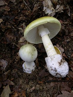
Amanita phalloides (generally pronounced /æməˈnaɪtə fəˈlɔɪdiːz/), commonly known as the death cap, is a poisonous basidiomycete fungus, one of many in the genus Amanita. Widely distributed across Europe, A. phalloides associates with various deciduous trees. In some cases, death cap has been accidentally introduced to new regions with the cultivation of non-native species of oak, chestnut, and pine. The large fruiting bodies (i.e., the mushrooms) appear in summer and autumn; the caps are generally greenish in colour, with a white stipe and gills.
Coincidentally, these toxic mushrooms resemble several edible species (most notably the straw mushroom) commonly consumed by humans, increasing the risk of accidental poisoning. A. phalloides is one of the most poisonous of all known toadstools. It has been involved in the majority of human deaths from mushroom poisoning,[1] possibly including the deaths of Roman Emperor Claudius and Holy Roman Emperor Charles VI. It has been the subject of much research, and many of its biologically active agents have been isolated. The principal toxic constituent is α-amanitin, which damages the liver and kidneys, often fatally. No antidote is known.
Contents |
Taxonomy and naming
The death cap was first described by French botanist Sébastien Vaillant in 1727, who gave a succinct phrase name "Fungus phalloides, annulatus, sordide virescens, et patulus", which is still recognizable as the fungus today.[2] Though the scientific name phalloides means "phallus-shaped", it is unclear whether it is named for its resemblance to a literal phallus or the stinkhorn mushrooms Phallus. In 1821, Elias Magnus Fries described it as Agaricus phalloides, but included all white Amanitas within its description.[3] Finally in 1833, Johann Heinrich Friedrich Link settled on the name Amanita phalloides,[4] after Persoon had named it Amanita viridis thirty years earlier.[5][6] Although Louis Secretan's use of the name Amanita phalloides predates Link's, it has been rejected for nomenclatural purposes because Secretan's works did not use binomial nomenclature consistently;[7][8] some taxonomists have, however, disagreed with this opinion.[9][10]
Amanita phalloides is the type species of Amanita section Phalloideae, a group that contains all of the deadly poisonous Amanita species thus far identified. Most notable of these are the species known as destroying angels, namely Amanita virosa and A. bisporiga as well as the fool's mushroom (A. verna). The term "destroying angel" has been applied to A. phalloides at times, but "death cap" is by far the most common vernacular name used in English. Other common names also listed include "stinking amanita"[11] and "deadly amanita".[12]
A rarely appearing all-white form was initially described A. phalloides f. alba by Max Britzelmayr,[13][14] though its status has been unclear. It is often found growing amid normally coloured death caps. It has been described, in 2004, as a distinct variety and includes what was termed A. verna var. tarda.[15] The true Amanita verna fruits in spring and turns yellow with KOH solution, whereas A. phalloides never does.[16]
Description
The death cap has a large and imposing epigeous (aboveground) fruiting body (basidiocarp), usually with a pileus (cap) from 5 to 15 cm (2–6 in) across, initially rounded and hemispherical, but flattening with age.[17] The colour of the cap can be pale-, yellowish-, or olive-green, often paler toward the margins and often paler after rain. The cap surface is sticky when wet and easily peeled, a troublesome feature, as that is allegedly a feature of edible fungi.[18] The remains of the partial veil is seen as a skirtlike, floppy annulus usually about 1 to 1.5 cm (0.4–0.6 in) below the cap. The crowded white lamellae (gills) are free. The stipe is white with a scattering of grayish-olive scales and is 8 to 15 cm (3–6 in) long and 1 to 2 cm (3/8–3/4 in) thick, with a swollen, ragged, sac-like white volva (base).[17] As the volva, which may be hidden by leaf litter, is a distinctive and diagnostic feature, it is important to remove some debris to check for it.[19]
The smell has been described as initially faint and honey-sweet but strengthening over time to become overpowering, sickly-sweet and objectionable.[20] Young specimens first emerge from the ground resembling a white egg covered by a universal veil, which then breaks, leaving the volva as a remnant. The spore print is white, a common feature of Amanita. The transparent spores are globular to egg-shaped, measure 8–10 μm (0.3–0.4 mil) long, and stain blue with iodine.[20] The gills, on the other hand, are seen to stain pallid lilac or pink with concentrated sulfuric acid.[21][22]
Distribution and habitat

The death cap is native to Europe, where it is widespread.[23] It is found from the southern coastal regions of Scandinavia in the north, to Ireland in the west, east to Poland and western Russia,[15] and south throughout the Balkans, in Italy and Spain, and in Morocco and Algeria in north Africa.[24] There are records from further east into Asia but these have yet to be confirmed as A. phalloides.[25]
It is ectomycorrhizally associated with a number of tree species. In Europe, these include a large number of hardwood and, less frequently, conifer species. It appears most commonly under oaks but also under beeches, chestnuts, horse-chestnuts, birches, filberts, hornbeams, pines, and spruces.[13] In other areas, A. phalloides may also be associated with these trees or only with some species but not others. In coastal California, for example, A. phalloides is associated with coast live oak but not with the various coastal pine species, such as Monterey pine.[26] In countries where it has been introduced it has been restricted to those exotic trees it would associate with in its natural range. There is, however, evidence of A. phalloides associating with hemlock and with genera of the Myrtaceae: Eucalyptus in Tanzania[27] and Algeria,[24] and Leptospermum and Kunzea in New Zealand.[28][13] This suggests the species may have invasive potential.[25]
By the end of the 19th century, Charles Horton Peck had reported A. phalloides in North America.[29] However, in 1918, samples from the Eastern United States were identified as being a distinct though similar species, A. brunnescens, by G. F. Atkinson of Cornell University.[30] By the 1970s it had become clear that A. phalloides actually does occur in the United States, apparently having been introduced from Europe alongside chestnuts, with populations on the West and East Coasts.[30][31] A more recent historical review concluded that the East Coast populations were introduced but that the origins of the West Coast population remain unclear, due to the scantness of historical records.[25]
Amanita phalloides has been conveyed to new countries across the southern hemisphere with the importation of hardwoods and conifers. Introduced oaks appear to have been the vector to Australia and South America; populations under oaks have been recorded from Melbourne and Canberra,[32][33] as well as Uruguay.[34] It has been recorded under other introduced trees in Argentina[35] and Chile.[36] Pine plantations are associated with the fungus in Tanzania[27] and South Africa, where it is also found under oaks and poplars.[37]
Toxicity

As the common name suggests, the fungus is highly toxic, and it is responsible for the majority of fatal mushroom poisonings worldwide.[1] It is estimated that 30 grams (1 oz), or half a cap, of this mushroom is enough to kill a human.[38] In 2006, a family of three in Poland was poisoned, resulting in one death and the two survivors requiring liver transplants.[39] Some authorities strongly advise against putting suspected death caps in the same basket with fungi collected for the table and to avoid touching them.[18][40] Furthermore, the toxicity is not reduced by cooking, freezing, or drying. Its biochemistry has been researched intensively for decades.[30]
Similarity to edible species
Recent cases highlight the issue of the similarity of A. phalloides to the edible paddy straw mushroom, Volvariella volvacea, with east- and southeast-Asian immigrants in Australia and the west coast of the United States falling victim. In an episode in Oregon, four members of a Korean family required liver transplants.[41] Of the seven people poisoned in the Canberra region between 1988 and 1998, three were from Laos.[42] This misidentification is a leading cause of mushroom poisoning in the United States.
Novices may mistake juvenile death caps for edible puffballs[43] or mature specimens for other edible Amanita species such as Amanita lanei, and for this reason some authorities recommend avoiding the collecting of Amanita species for the table altogether.[44] The white form of A. phalloides may be mistaken for edible species of Agaricus, especially the young fruitbodies whose unexpanded caps conceal the telltale white gills; all mature species of Agaricus have dark-coloured gills.[45]
In Europe, other similarly green-capped species collected by mushroom hunters include various green-hued brittlegills of the genus Russula and the formerly popular Tricholoma flavovirens, now regarded as hazardous owing to a series of restaurant poisonings in France. Brittlegills, such as Russula heterophylla, R. aeruginea, and R. virescens, can be distinguished by their brittle flesh and the lack of both volva and ring.[46] Other similar species include A. subjunquillea in eastern Asia and A. arocheae, which ranges from Andean Colombia north at least as far as central Mexico, both of which are also poisonous.
Biochemistry
The species is now known to contain two main groups of toxins, both multicyclic (ring-shaped) peptides, spread throughout the mushroom tissue: the amatoxins and the phallotoxins. Another toxin is phallolysin, which has shown some hemolytic (red blood cell–destroying) activity in vitro. An unrelated compound, antamanide, has also been isolated.
Amatoxins consist of at least eight compounds with a similar structure, that of eight amino-acid rings; they were isolated in 1941 by Heinrich O. Wieland and Rudolf Hallermayer of the University of Munich.[30] Of the amatoxins, α-amanitin is the chief component and along with β-amanitin is likely responsible for the toxic effects.[47][48] Their major toxic mechanism is the inhibition of RNA polymerase II, a vital enzyme in the synthesis of messenger RNA (mRNA), microRNA, and small nuclear RNA (snRNA). Without mRNA essential protein synthesis and hence cell metabolism grind to a halt and the cell dies.[49] The liver is the principal organ affected, as it is the organ which is first encountered after absorption in the gastrointestinal tract, though other organs, especially the kidneys, are susceptible.[50] The RNA polymerase of Amanita phalloides is insensitive to the effects of amatoxins; as such, the mushroom does not poison itself.[51]
The phallotoxins consist of at least seven compounds, all of which have seven similar peptide rings. Phalloidin was isolated in 1937 by Feodor Lynen, Heinrich Wieland's student and son-in-law, and Ulrich Wieland of the University of Munich. Though phallotoxins are highly toxic to liver cells,[52] they have since been found to have little input into the death cap's toxicity as they are not absorbed through the gut.[49] Furthermore, phalloidin is also found in the edible (and sought-after) Blusher (Amanita rubescens).[30] Another group of minor active peptides are the virotoxins, which consist of six similar monocyclic heptapeptides.[53] Like the phallotoxins they do not exert any acute toxicity after ingestion in humans.[49]
Symptoms
Death caps have been reported to taste pleasant.[30][54] This, coupled with the delay in the appearance of symptoms—during which time internal organs are being severely, sometimes irreparably, damaged—makes it particularly dangerous. Initially, symptoms are gastrointestinal in nature and include colicky abdominal pain, with watery diarrhea and vomiting which may lead to dehydration, and, in severe cases, hypotension, tachycardia, hypoglycemia, and acid-base disturbances.[55][56] These first symptoms resolve two to three days after the ingestion. A more serious deterioration signifying liver involvement may then occur—jaundice, diarrhea, delirium, seizures, and coma due to fulminant hepatic failure and attendant hepatic encephalopathy caused by the accumulation of normally liver-removed substance in the blood.[11] Renal failure (either secondary to severe hepatitis[57][53] or caused by direct toxic renal damage[49]) and coagulopathy may appear during this stage. Life-threatening complications include increased intracranial pressure, intracranial hemorrhage, sepsis, pancreatitis, acute renal failure, and cardiac arrest.[55][56] Death generally occurs six to sixteen days after the poisoning.[58]
Up to the mid-20th century, the mortality rate was around 60–70%, but this has greatly improved with advances in medical care. A review of death cap poisoning throughout Europe from 1971 to 1980 found the overall mortality rate to be 22.4% (51.3% in children under ten and 16.5% in those older than ten).[59] This has fallen further in more recent surveys to around 10–15%.[60]
Treatment
Consumption of the death cap is a medical emergency requiring hospitalization. There are four main categories of therapy for poisoning: preliminary medical care, supportive measures, specific treatments, and liver transplantation.[61]
Preliminary care consists of gastric decontamination with either activated carbon or gastric lavage. However, due to the delay between ingestion and the first symptoms of poisoning, it is commonplace for patients to arrive for treatment many hours after ingestion, potentially reducing the efficacy of these interventions.[61][62] Supportive measures are directed towards treating the dehydration which results from fluid loss during the gastrointestinal phase of intoxication and correction of metabolic acidosis, hypoglycemia, electrolyte imbalances, and impaired coagulation.[61]
No definitive antidote is available, but some specific treatments have been shown to improve survivability. High-dose continuous intravenous penicillin G has been reported to be of benefit, though the exact mechanism is unknown,[59] and trials with cephalosporins show promise.[63][64] There is some evidence that intravenous silibinin, an extract from the blessed milk thistle (Silybum marianum), may be beneficial in reducing the effects of death cap poisoning. Silibinin prevents the uptake of amatoxins by hepatocytes, thereby protecting undamaged hepatic tissue; it also stimulates DNA-dependent RNA polymerases, leading to an increase in RNA synthesis.[65][66][67] N-acetylcysteine has shown promise in combination with other therapies.[68] Animal studies indicate the amatoxins deplete hepatic glutathione;[69] N-acetylcysteine serves as a glutathione precursor and may therefore prevent reduced glutathione levels and subsequent liver damage.[70] None of the antidotes used have undergone prospective, randomized clinical trials, and only anecdotal support is available. Silibinin and N-acetylcysteine appear to be the therapies with the most potential benefit.[61] Repeated doses of activated carbon may be helpful by absorbing any toxins that are returned to the gastrointestinal tract following enterohepatic circulation.[71] Other methods of enhancing the elimination of the toxins have been trialed; techniques such as hemodialysis,[72] hemoperfusion,[73] plasmapheresis,[74] and peritoneal dialysis[75] have occasionally yielded success but overall do not appear to improve outcome.[49]
In patients developing liver failure, a liver transplant is often the only option to prevent death. Liver transplants have become a well-established option in amatoxin poisoning.[56][55][76] This is a complicated issue, however, as transplants themselves may have significant complications and mortality; patients require long-term immunosuppression to maintain the transplant.[61] That being the case, there has been a reassessment of criteria such as onset of symptoms, prothrombin time (PTT), serum bilirubin, and presence of encephalopathy for determining at what point a transplant becomes necessary for survival.[77][78][79] Evidence suggests that, although survival rates have improved with modern medical treatment, in patients with moderate to severe poisoning up to half of those who did recover suffered permanent liver damage.[80] However, a follow-up study has shown that most survivors recover completely without any sequelae if treated within 36 hours of mushroom ingestion.[81]
Notable victims
Several historical figures may have died from Amanita phalloides poisoning (or other similar, toxic Amanitas). These were either accidental poisonings or assassination plots. Alleged victims of this kind of poisoning include Roman Emperor Claudius, Pope Clement VII, Tsaritsa Natalia Naryshkina, and Holy Roman Emperor Charles VI.[82]
R. Gordon Wasson recounted the details of these deaths, noting the likelihood of Amanita poisoning. In the case of Clement VII, the illness that led to his death lasted some five months, making the case clearly inconsistent with amatoxin poisoning. Natalia Naryshkina is said to have consumed a large quantity of pickled mushrooms prior to her death. However, it is unclear whether the mushrooms themselves were poisonous or whether she succumbed to food poisoning.[82]
| “ | Ce plat de champignons a changé la destinée de l’Europe. ("This dish of mushrooms changed the destiny of Europe.") |
” |
|
—Voltaire, Mémoires. |
||
Charles VI experienced indigestion after eating a dish of sautéed mushrooms. This led to an illness from which he died ten days later — symptomology consistent with amatoxin poisoning. Charles' death led to the War of Austrian Succession. Noted Voltaire, "this dish of mushrooms changed the destiny of Europe."[83][82]
The case of the Claudius poisoning is more complex. It is known that Claudius was very fond of eating Caesar's mushroom. Following his death, many sources have attributed it to his being fed a meal of death caps instead of Caesar's mushrooms. However, ancient authors such as Tacitus and Suetonius are unanimous about there having been poison added to the mushroom dish, rather than the dish having been prepared from poisonous mushrooms. Wasson speculates that the poison used to kill Claudius was derived from death caps, with a fatal dose of colocynth being administered later during his illness.[84][82]
Footnotes
- ↑ 1.0 1.1 Benjamin.p200
- ↑ (Latin)Vaillant, Sébastien (1727). Botanicon Parisiense. Leide & Amsterdam: J. H. Verbeek and B. Lakeman. OCLC 5146641.
- ↑ (Latin)Fries, Elias Magnus (1821). Systema Mycologicum I. Gryphiswaldiae: Ernesti Mauritii. OCLC 10717479.
- ↑ (German) Link JHF (1833) Grundriss der Kraeuterkunde IV. Haude und Spenerschen Buchhandlung (S.J. Joseephy), Berlin
- ↑ (Latin)Persoon, Christian Hendrik (1797). Tentamen Dispositionis Methodicae Fungorum. Lipsiae: P.P. Wolf,. OCLC 19300194.
- ↑ (Latin)Persoon, Christian Hendrik (1801). Synopsis Methodica Fungorum. Göttingen: H. Dietrich. OCLC 28329773.
- ↑ Donk, M.A. (June 1962). "On Secretan's Fungus Names". Taxon 11 (5): 170–173. doi:. http://links.jstor.org/sici?sici=0040-0262%28196206%2911%3A5%3C170%3AOSFN%3E2.0.CO%3B2-I.
- ↑ Demoulin, V. (November 1974). "Invalidity of Names Published in Secretan's Mycographie Suisse and Some Remarks on the Problem of Publication by Reference". Taxon 23 (5/6): 836–843. doi:. http://links.jstor.org/sici?sici=0040-0262%28197411%2923%3A5%2F6%3C836%3AIONPIS%3E2.0.CO%3B2-T.
- ↑ Singer, Rolf; Robert E. Machol (June 1962). "Are Secretan's Fungus Names Valid?". Taxon 26 (2/3): 251–255. doi:. http://links.jstor.org/sici?sici=0040-0262%28197705%2926%3A2%2F3%3C251%3AASFNV%3E2.0.CO%3B2-G.
- ↑ Machol, Robert E. (August 1984). "Leave the Code Alone". Taxon 33 (3): 532–533. doi:. http://links.jstor.org/sici?sici=0040-0262%28198408%2933%3A3%3C532%3ALTCA%3E2.0.CO%3B2-9.
- ↑ 11.0 11.1 North, Pamela Mildred (1967). Poisonous plants and fungi in colour. London: Blandford Press. OCLC 955264.
- ↑ Benjamin.p203
- ↑ 13.0 13.1 13.2 Tulloss, Rodham E.. "(Fr.:Fr.) Link". Amanita Studies site. Retrieved on 2007-05-22.
- ↑ Jordan & Wheeler. p109
- ↑ 15.0 15.1 Neville, Pierre; Serge Poumarat (2004). Amaniteae: Amanita, Limacella and Torrendia'. Fungi Europaei (9). Alassio. ISBN 88-901057-3-9.
- ↑ Tulloss, Rodham E.. "Amanita verna (Bull.: Fr.) Lam.". Amanita Studies site. Retrieved on 2007-05-22.
- ↑ 17.0 17.1 Bresinsky A, Besl H. (1990). A Colour Atlas of Poisonous Fungi. Wolfe Publishing. pp. 26-9. ISBN 0-7234-1576-5.
- ↑ 18.0 18.1 Jordan & Wheeler. p99
- ↑ Jordan & Wheeler. p108
- ↑ 20.0 20.1 Zeitlmayr. p61
- ↑ Jordan, Michael (1995). The Encyclopedia of Fungi of Britain and Europe. David & Charles. pp. 198. ISBN 0-7153-0129-2.
- ↑ "California Fungi: Amanita phalloides". MykoWeb.com. Retrieved on June 01, 2007.
- ↑ Lange, Lene (1974). "The distribution of macromycetes in Europe". Dansk Botanisk Arkiv 30: 5–105. ISSN 0011-6211.
- ↑ 24.0 24.1 Malençon, Georges; R. Bertault (1970). Flore des Champignons Supérieurs du Maroc I. Travaux de l'Institut scientifique chérifien et de la Faculté des sciences. Série botanique et biologie végétale (32). Rabat: Faculté des Sciences. OCLC 915096.
- ↑ 25.0 25.1 25.2 Pringle, Anne; Else C. Vellinga (July 2006). "Last chance to know? Using literature to explore the biogeography of and invasion biology of the death cap mushroom Amanita phalloides (Vaill. Ex Fr. :Fr) Link". Biological Invasions 8 (5): 1131–1144. doi:.
- ↑ Arora, David (1986). Mushrooms demystified : a comprehensive guide to the fleshy fungi. Berkeley, California: Ten Speed Press. ISBN 0-89815-170-8.
- ↑ 27.0 27.1 Pegler (1977). A preliminary agaric flora of East Africa. Kew Bulletin Additional Series (6). Royal Botanic Gardens, Kew. ISBN 0-11-241101-0.
- ↑ Ridley, G.S. (1991). "The New Zealand Species of Amanita (Fungi: Agaricales)". Australian Systematic Botany 4 (2): 325–354. doi:.
- ↑ Peck, Charles H. (1897). Annual report of the state botanist. Albany: University of the State of New York. OCLC 1185748.
- ↑ 30.0 30.1 30.2 30.3 30.4 30.5 Litten, W. (March 1975). "The most poisonous mushrooms". Scientific American 232 (3): 90–101. PMID 1114308.
- ↑ Benjamin.p204
- ↑ Reid, D.A. (1980). "A monograph of the Australian species of Amanita Pers. ex Hook (Fungi)". Australian Journal of Botany Supplementary Series 8: 1–96.
- ↑ Cole, F.M. (June 1993). "Amanita phalloides in Victoria". Medical Journal of Australia 158 (12): 849–850. PMID 8326898.
- ↑ (Spanish)Herter, W.G. (1934). "La aparición del hongo venenoso Amanita phalloides en Sudamérica.". Revista Sudamericana de Botánica 1: 111–119.
- ↑ (Spanish)Hunzinker, A.T. (1983). "Amanita phalloides en las Sierras de Córdoba". Kurtziana 16: 157–160. ISSN 0075-7314.
- ↑ Valenzuella, E.; G. Moreno & M. Jeria (1992). "Amanita phalloides en bosques de Pinus radiata de la IX Region de Chile: taxonomia, toxinas, metodos de dedection, intoxicacion faloidiana". Boletín Micológico 7: 17–21. ISSN 0716-114X.
- ↑ Reid, D.A.; A. Eicker (1991). "South African fungi: the genus Amanita". Mycological Research 95 (1): 80–95. ISSN 0953-7562.
- ↑ Benjamin.p211
- ↑ (Polish) Pawlowska, J.; Pawlak J, Kamiski A, Hevelke P, Jankowska I, Teisseyre M, Szymczak M, Kaliciiski P, Krawczyk M. (2006). "(Amanita phalloides poisoning as an indication for liver transplantation in three family members.)". Wiadomości Lekarskie 59 (1–2): 131–4. PMID 16646310.
- ↑ Carluccio A (2003). The Complete Mushroom Book. London: Quadrille. pp. 224. ISBN 1-84400-040-0.
- ↑ Benjamin.p198–199
- ↑ Trim Geoffrey M. et al. (September 1999). "Poisoning by Amanita phalloides ("deathcap") mushrooms in the Australian Capital Territory". Medical Journal of Australia 171 (5): 247–249. PMID 10495756. http://www.mja.com.au/public/issues/171_5_060999/trim/trim.html. Retrieved on 2007-05-22.
- ↑ Hall IR, Stephenson SE, Buchanan PK, Yn W, Cole AL. (2003). Edible and poisonous mushrooms of the world. New Zealand Institute for Crop & Food Research Limited. pp. 131-3. ISBN 0-4781-0835-4.
- ↑ Phillips, Roger (2005). Mushrooms and Other Fungi of North America. Buffalo: Firefly books. pp. p.14. ISBN 1-55407-115-1.
- ↑ Heino, Lepp (2006-10-09). "Deathcap Mushroom: Amanita phalloides". Australian National Botanic Gardens. Retrieved on 2007-06-12.
- ↑ Zeitlmayr. p62
- ↑ Köppel C (1993). "Clinical symptomatology and management of mushroom poisoning". Toxicon 31 (12): 1513–40. doi:. PMID 8146866.
- ↑ Dart, RC (2004). "Mushrooms". Medical toxicology. Philadelphia: Williams & Wilkins. pp. 1719–35. ISBN 0-7817-2845-2.
- ↑ 49.0 49.1 49.2 49.3 49.4 Karlson-Stiber C, Persson H (2003). "Cytotoxic fungi - an overview". Toxicon 42 (4): 339–49. doi:. PMID 14505933.
- ↑ Benjamin.p217
- ↑ Horgen, Paul A.; Allan C. Vaisius and Joseph F. Ammirati (1978). "The insensitivity of mushroom nuclear RNA polymerase activity to inhibition by amatoxins". Archives of Microbiology 118 (3): 317–9. doi:. PMID 567964.
- ↑ Wieland T, Govindan VM (1974). "Phallotoxins bind to actins". FEBS Lett. 46 (1): 351–3. doi:. PMID 4429639.
- ↑ 53.0 53.1 Vetter, János (January 1998). "Toxins of Amanita phalloides". Toxicon 36 (1): 13–24. doi:. PMID 9604278.
- ↑ Cleland, John Burton (1976) [1934]. Toadstools and mushrooms and other larger fungi of South Australia. South Australian Government Printer. OCLC 15150059.
- ↑ 55.0 55.1 55.2 Pinson CW, Daya MR, Benner KG, Norton RL, Deveney KE, Ascher NL, Roberts JP, Lake JR, Kurkchubasche AG, Ragsdale JW (May 1990). "Liver transplantation for severe Amanita phalloides mushroom poisoning". American Journal of Surgery 159 (5): 493–9. doi:. PMID 2334013.
- ↑ 56.0 56.1 56.2 Klein AS, Hart J, Brems JJ, Goldstein L, Lewin K, Busuttil RW (February 1989). "Amanita poisoning: treatment and the role of liver transplantation". American Journal of Medicine 86 (2): 187–93. doi:. PMID 2643869.
- ↑ Nicholls DW, Hyne BE, Buchanan P (1995). "Death cap mushroom poisoning". The New Zealand Medical Journal 108 (1001): 234. PMID 7603660.
- ↑ Fineschi V, Di Paolo M, Centini F (1996). "Histological criteria for diagnosis of amanita phalloides poisoning". J. Forensic Sci. 41 (3): 429–32. PMID 8656182.
- ↑ 59.0 59.1 (German)Floerscheim, G.L.; O. Weber, P. Tschumi & M. Ulbrich (August 1982). "Die klinische knollenblatterpilzvergiftung (Amanita Phalloides): prognostische faktoren und therapeutische massnahmen (Clinical death-cap (Amanita phalloides) poisoning: prognostic factors and therapeutic measures.)". Schweizerische medizinische Wochenschrift 112 (34): 1164–1177. PMID 6291147.
- ↑ Benjamin.p215
- ↑ 61.0 61.1 61.2 61.3 61.4 Enjalbert F, Rapior S, Nouguier-Soulé J, Guillon S, Amouroux N, Cabot C (2002). "Treatment of amatoxin poisoning: 20-year retrospective analysis". Journal of Toxicology - Clinical Toxicology 40 (6): 715–57. PMID 12475187.
- ↑ Vesconi S, Langer M, Iapichino G, Costantino D, Busi C, Fiume L (1985). "Therapy of cytotoxic mushroom intoxication". Critical care medicine 13 (5): 402–6. doi:. PMID 3987318.
- ↑ Benjamin.p227
- ↑ (German)Neftel, K. et al. (January 1988). "(Are cephalosporins more active than penicillin G in poisoning with the deadly Amanita?)". Schweizerische medizinische Wochenschrift 118 (2): 49–51. PMID 3278370.
- ↑ Hruby K, Csomos G, Fuhrmann M, Thaler H (1983). "Chemotherapy of Amanita phalloides poisoning with intravenous silibinin". Human toxicology 2 (2): 183–95. PMID 6862461.
- ↑ (Italian) Carducci, R. et al. (May 1996). "[[Silibinin and acute poisoning with Amanita phalloides]]". Minerva Anestesiologica 62 (5): 187–93. PMID 8937042.
- ↑ Jahn, W. (1980). "Pharmacokinetics of {3H}-methyl-dehydroxymethyl-amanitin in the isolated perfused rat liver, and the influence of several drugs". in Helmuth Faulstich, B. Kommerell & Theodore Wieland. Amanita toxins and poisoning. Baden-Baden: Witzstrock. pp. 80–85. ISBN 3-87921-132-9.
- ↑ Montanini S, Sinardi D, Praticò C, Sinardi A, Trimarchi G (1999). "Use of acetylcysteine as the life-saving antidote in Amanita phalloides (death cap) poisoning. Case report on 11 patients". Arzneimittel-Forschung 49 (12): 1044–7. PMID 10635453.
- ↑ Kawaji A, Sone T, Natsuki R, Isobe M, Takabatake E, Yamaura Y (1990). "In vitro toxicity test of poisonous mushroom extracts with isolated rat hepatocytes". The Journal of toxicological sciences 15 (3): 145–56. PMID 2243367.
- ↑ Chyka P, Butler A, Holliman B, Herman M (2000). "Utility of acetylcysteine in treating poisonings and adverse drug reactions". Drug safety 22 (2): 123–48. doi:. PMID 10672895.
- ↑ Busi C, Fiume L, Costantino D, Langer M, Vesconi F (1979). "Amanita toxins in gastroduodenal fluid of patients poisoned by the mushroom, Amanita phalloides". New England Journal of Medicine 300 (14): 800. PMID 423916.
- ↑ Sabeel AI, Kurkus J, Lindholm T (1995). "Intensive hemodialysis and hemoperfusion treatment of Amanita mushroom poisoning". Mycopathologia 131 (2): 107–14. doi:. PMID 8532053.
- ↑ Wauters JP, Rossel C, Farquet JJ (1978). "Amanita phalloides poisoning treated by early charcoal haemoperfusion". British medical journal 2 (6150): 1465. PMID 719466.
- ↑ Jander S, Bischoff J, Woodcock BG (2000). "Plasmapheresis in the treatment of Amanita phalloides poisoning: II. A review and recommendations". Therapeutic apheresis 4 (4): 308–12. doi:. PMID 10975479.
- ↑ Langer M, Vesconi S, Iapichino G, Costantino D, Radrizzani D (1980). "The early removal of amatoxins in the treatment of amanita phalloides poisoning" (in German). Klinische Wochenschrift 58 (3): 117–23. doi:. PMID 7366125.
- ↑ Ganzert M, Felgenhauer N, Zilker T (2005). "Indication of liver transplantation following amatoxin intoxication". Journal of Hepatology 42 (2): 202–9. doi:. PMID 15664245.
- ↑ O'grady, John G.; Graeme J.M. Alexander, Karen M. Hayllar & Roger Williams (august 1989). "Early indicators of prognosis in fulminant hepatic failure". Gastroenterology 97 (2): 439–445. PMID 2490426.
- ↑ Panaro, Fabrizio; Enzo Andorno, Nicola Morelli, Marco Casaccia, Giuliano Bottino, Ferruccio Ravazzoni, Monica Centanaro, Sara Ornis & Umberto Valente (April 2006). "Letter to the editor: Liver transplantation represents the optimal treatment for fulminant hepatic failure from Amanita phalloides poisoning". Transplant International 19 (4): 344–5. doi:. PMID 16573553.
- ↑ Escudié L, Francoz C, Vinel JP, Moucari R, Cournot M, Paradis V, Sauvanet A, Belghiti J, Valla D, Bernuau J, Durand F (2007). "Amanita phalloides poisoning: reassessment of prognostic factors and indications for emergency liver transplantation". J. Hepatol. 46 (3): 466–73. doi:. PMID 17188393.
- ↑ Benjamin.p231–232
- ↑ Giannini L, Vannacci A, Missanelli A, Mastroianni R, Mannaioni PF, Moroni F, Masini E (2007). "Amatoxin poisoning: A 15-year retrospective analysis and follow-up evaluation of 105 patients". Clinical toxicology (Philadelphia, Pa.) 45 (5): 539–42. doi:. PMID 17503263.
- ↑ 82.0 82.1 82.2 82.3 Wasson, Robert Gordon (1972). "The death of Claudius, or mushrooms for murderers". Botanical Museum Leaflets, Harvard University 23 (3): 101–128. ISSN 0006-8098.
- ↑ Benjamin.p35
- ↑ Benjamin.p33–34
Sources
- Benjamin, Denis R. (1995). Mushrooms: poisons and panaceas — a handbook for naturalists, mycologists and physicians. New York: WH Freeman and Company. ISBN 0-7167-2600-9.
- Jordan Peter, Wheeler Steven. (2001). The Ultimate Mushroom Book. London: Hermes House. ISBN 1-85967-092-X.
- Zeitlmayr, Linus (1976). Wild Mushrooms:An Illustrated Handbook. Hertfordshire: Garden City Press. ISBN 0-584-10324-7.
External links
- UK Telegraph Newspaper (Sept 2008) - One woman dead, another critically ill after eating Death Cap fungi
- AmericanMushrooms.com - The Death Cap Mushroom Amanita phalloides
- Amanita phalloides: the death cap
- Key to species of Amanita Section Phalloidieae from North and Central America - Amanita studies website
- Death cap in Australia - ANBG website
- On the Trail of the Death Cap Mushroom from American National Public Radio
- Amanita phalloides at the Encyclopedia of Life